

Nanomaterials for Skin Cancer Photoimmunotherapy
- PMID: 37238966
- PMCID: PMC10215838
- DOI: 10.3390/biomedicines11051292
Nanomaterials for Skin Cancer Photoimmunotherapy
Abstract
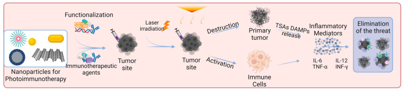
Skin cancer is one of the most common types of cancer, and its incidence continues to increase. It is divided into two main categories, melanoma and non-melanoma. Treatments include surgery, radiation therapy, and chemotherapy. The relatively high mortality in melanoma and the existing recurrence rates, both for melanoma and non-melanoma, create the need for studying and developing new approaches for skin cancer management. Recent studies have focused on immunotherapy, photodynamic therapy, photothermal therapy, and photoimmunotherapy. Photoimmunotherapy has gained much attention due to its excellent potential outcomes. It combines the advantages of photodynamic and/or photothermal therapy with a systemic immune response, making it ideal for metastatic cancer. This review critically discusses different new nanomaterials' properties and mechanisms of action for skin cancer photoimmunotherapy and the main results obtained in the field.
Keywords: basal cell carcinoma; immunotherapy; melanoma; photodynamic therapy; photothermal therapy; squamous cell carcinoma.
Conflict of interest statement
The authors declare no conflict of interest.
Figures


Similar articles
-
Multifunctional inorganic nanomaterials for cancer photoimmunotherapy.Cancer Commun (Lond). 2022 Feb;42(2):141-163. doi: 10.1002/cac2.12255. Epub 2022 Jan 9. Cancer Commun (Lond). 2022. PMID: 35001556 Free PMC article. Review.
-
Combination of PEG-decorated black phosphorus nanosheets and immunoadjuvant for photoimmunotherapy of melanoma.J Mater Chem B. 2020 Apr 8;8(14):2805-2813. doi: 10.1039/d0tb00434k. J Mater Chem B. 2020. PMID: 32163088
-
Combination of NIR therapy and regulatory T cell modulation using layer-by-layer hybrid nanoparticles for effective cancer photoimmunotherapy.Theranostics. 2018 Aug 10;8(17):4574-4590. doi: 10.7150/thno.26758. eCollection 2018. Theranostics. 2018. PMID: 30279723 Free PMC article.
-
Recent Advances on Rare Earth Upconversion Nanomaterials for Combined Tumor Near-Infrared Photoimmunotherapy.Front Chem. 2020 Nov 6;8:596658. doi: 10.3389/fchem.2020.596658. eCollection 2020. Front Chem. 2020. PMID: 33240857 Free PMC article. Review.
-
Antibody-Based Immunotherapy: Alternative Approaches for the Treatment of Metastatic Melanoma.Biomedicines. 2020 Sep 3;8(9):327. doi: 10.3390/biomedicines8090327. Biomedicines. 2020. PMID: 32899183 Free PMC article. Review.
Cited by
-
Phase-specific and laser-modulated polydopamine-chlorella-curdlan hydrogels: pioneering a melanoma integrative therapy.Theranostics. 2025 Jun 23;15(15):7627-7652. doi: 10.7150/thno.113417. eCollection 2025. Theranostics. 2025. PMID: 40756368 Free PMC article.
-
Advances in Materials Science for Precision Melanoma Therapy: Nanotechnology-Enhanced Drug Delivery Systems.Pharmaceutics. 2025 Feb 24;17(3):296. doi: 10.3390/pharmaceutics17030296. Pharmaceutics. 2025. PMID: 40142960 Free PMC article. Review.
References
-
- Craythorne E., Al-Niami F. Skin cancer. Medicine. 2017;45:431–434. doi: 10.1016/j.mpmed.2017.04.003. - DOI
-
- World Health Organization International Agency for Research on Cancer GLOBOCAN 2020: Estimated Cancer Incidence, Mortality and Prevalence Worldwide in 2020. [(accessed on 10 April 2023)]. Available online: https://gco.iarc.fr/today/online-analysis-pie?v=2020&mode=population&mod....
Publication types
Grants and funding
LinkOut - more resources
Full Text Sources
