. 2023 May 4;11(5):1355.
doi: 10.3390/biomedicines11051355.

Comparison of the Penetration Depth of 905 nm and 1064 nm Laser Light in Surface Layers of Biological Tissue Ex Vivo

Affiliations

Comparison of the Penetration Depth of 905 nm and 1064 nm Laser Light in Surface Layers of Biological Tissue Ex Vivo

Leon Kaub et al. Biomedicines. .

Abstract

The choice of parameters for laser beams used in the treatment of musculoskeletal diseases is of great importance. First, to reach high penetration depths into biological tissue and, secondly, to achieve the required effects on a molecular level. The penetration depth depends on the wavelength since there are multiple light-absorbing and scattering molecules in tissue with different absorption spectra. The present study is the first comparing the penetration depth of 1064 nm laser light with light of a smaller wavelength (905 nm) using high-fidelity laser measurement technology. Penetration depths in two types of tissue ex vivo (porcine skin and bovine muscle) were investigated. The transmittance of 1064 nm light through both tissue types was consistently higher than of 905 nm light. The largest differences (up to 5.9%) were seen in the upper 10 mm of tissue, while the difference vanished with increasing tissue thickness. Overall, the differences in penetration depth were comparably small. These results may be of relevance in the selection of a certain wavelength in the treatment of musculoskeletal diseases with laser therapy.

Keywords: 1064 nm NIR laser; 905 nm NIR laser; continuous wave lasers; laser therapy; musculoskeletal system; porcine/bovine tissues; pulsed lasers; tissue penetration depth.


Conflict of interest statement

Up until December 2017, C.S. served as a consultant for Electro Medical Systems (Nyon, Switzerland), and has received funding from Electro Medical Systems for conducting basic research on radial extracorporeal shock wave therapy and laser therapy at his lab. Regarding the present study, Electro Medical Systems did not impose any constraints on publication of the data and had no role in the design of the study; in data collection, analyses, or interpretation; in the writing of the manuscript; or in the decision to publish the findings.

Figures

Figure 1
Figure 1
Equipment used in the present study: (a) experimental setup of penetration depth measurements showing both the BTL laser (BTL) and the EMS laser (EMS); the thermal power sensor (sensor) in its location for penetration depth measurements as well as the porcine tissue specimens (red arrow) are also shown; (b) thermal power sensor; (c) photodiode sensor connected to an oscilloscope measuring pulses emitted by the EMS laser; and (d) beam profiling camera. Details are in the text. Panels (bd) were modified from [8] with permission from the authors.
Figure 2
Figure 2
Power measurements of the BTL laser: (a,b) measured average power (Pm) for different power values set at the BTL laser (Pset). Black dashed lines in (a,b) illustrate ideal curves, in which the measured average power would equal the set power; (c,d) deviation in percent of Pm compared to Pset. Measurements are shown for continuous-wave (CW) mode (a,c) and for pulsed-wave (PW) mode at three different repetition rates (b,d).
Figure 3
Figure 3
Characterization of the laser beam emitted by the BTL laser: (a) temporal profiles at different frequencies and the same peak power (12 W); (b) temporal profiles at the same frequency (100 Hz) at different values of peak power; (c) spatial intensity distribution; and (d) beam profiles along the horizontal line for different set beam sizes (zero indicates the center of the spatial intensity profile shown in (c)).
Figure 4
Figure 4
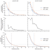
Penetration curves and differences in transmittance of the two LTDs (EMS laser, blue curves; BTL laser, orange curves) for the two tissues investigated: (a,c,e) porcine skin tissue; and (b,d,f) bovine muscle tissue. Transmittance was plotted against specimen thickness for the uncorrected data (a,b), the difference between the two LTDs in the uncorrected transmittance (c,d) and the corrected data (e,f). The 10% penetration depths are visualized in (a,b,e,f) by vertical dashed lines in the color of the LTD. The standard deviation of the measurements at each data point is given in (a,b) by vertical lines.
Figure 5
Figure 5
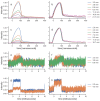
Recordings of laser pulses transmitted through tissue specimens: (a,b) EMS laser through porcine tissue; (c,d) EMS laser through bovine tissue; (e,f) BTL laser through porcine tissue; and (g,h) BTL laser through bovine tissue. The recordings were normalized in two ways: (a,c,e,g) normalized to the overall maximum of each laser therapy device and tissue; and (b,d,f,h) normalized to the individual maximum of each signal. All signals were smoothed using a moving average with a window length of 100 samples.

References

    1. Naterstad I.F., Joensen J., Bjordal J.M., Couppé C., Lopes-Martins R.A.B., Stausholm M.B. Efficacy of low-level laser therapy in patients with lower extremity tendinopathy or plantar fasciitis: Systematic review and meta-analysis of randomised controlled trials. BMJ Open. 2022;12:e059479. doi: 10.1136/bmjopen-2021-059479. - DOI - PMC - PubMed
    1. Mussttaf R.A., Jenkins D.F.L., Jha A.N. Assessing the impact of low level laser therapy (LLLT) on biological systems: A review. Int. J. Radiat. Biol. 2019;95:120–143. doi: 10.1080/09553002.2019.1524944. - DOI - PubMed
    1. Bjordal J.M., Lopes-Martins R.A.B., Joensen J., Iversen V.V. The anti-inflammatory mechanism of low level laser therapy and its relevance for clinical use in physiotherapy. Phys. Ther. Rev. 2010;15:286–293. doi: 10.1179/1743288X10Y.0000000001. - DOI
    1. Dompe C., Moncrieff L., Matys J., Grzech-Leśniak K., Kocherova I., Bryja A., Bruska M., Dominiak M., Mozdziak P., Skiba T.H.I., et al. Photobiomodulation—Underlying mechanism and clinical applications. J. Clin. Med. 2020;9:1724. doi: 10.3390/jcm9061724. - DOI - PMC - PubMed
    1. Schmitz C. Improving extracorporeal shock wave therapy with 904 or 905 nm pulsed, high power laser pretreatment. Preprints. 2021:2021010138. doi: 10.20944/preprints202101.0138.v1. - DOI

LinkOut - more resources