. 2023 May 5;11(5):1375.
doi: 10.3390/biomedicines11051375.

STING-Triggered CNS Inflammation in Human Neurodegenerative Diseases

Affiliations

STING-Triggered CNS Inflammation in Human Neurodegenerative Diseases

Alex S Ferecskó et al. Biomedicines. .

Abstract

Background: Some neurodegenerative diseases have an element of neuroinflammation that is triggered by viral nucleic acids, resulting in the generation of type I interferons. In the cGAS-STING pathway, microbial and host-derived DNA bind and activate the DNA sensor cGAS, and the resulting cyclic dinucleotide, 2'3-cGAMP, binds to a critical adaptor protein, stimulator of interferon genes (STING), which leads to activation of downstream pathway components. However, there is limited work demonstrating the activation of the cGAS-STING pathway in human neurodegenerative diseases.

Methods: Post-mortem CNS tissue from donors with multiple sclerosis (n = 4), Alzheimer's disease (n = 6), Parkinson's disease (n = 3), amyotrophic lateral sclerosis (n = 3) and non-neurodegenerative controls (n = 11) were screened by immunohistochemistry for STING and relevant protein aggregates (e.g., amyloid-β, α-synuclein, TDP-43). Human brain endothelial cells were cultured and stimulated with the STING agonist palmitic acid (1-400 μM) and assessed for mitochondrial stress (release of mitochondrial DNA into cytosol, increased oxygen consumption), downstream regulator factors, TBK-1/pIRF3 and inflammatory biomarker interferon-β release and changes in ICAM-1 integrin expression.

Results: In neurodegenerative brain diseases, elevated STING protein was observed mainly in brain endothelial cells and neurons, compared to non-neurodegenerative control tissues where STING protein staining was weaker. Interestingly, a higher STING presence was associated with toxic protein aggregates (e.g., in neurons). Similarly high STING protein levels were observed within acute demyelinating lesions in multiple sclerosis subjects. To understand non-microbial/metabolic stress activation of the cGAS-STING pathway, brain endothelial cells were treated with palmitic acid. This evoked mitochondrial respiratory stress up to a ~2.5-fold increase in cellular oxygen consumption. Palmitic acid induced a statistically significant increase in cytosolic DNA leakage from endothelial cell mitochondria (Mander's coefficient; p < 0.05) and a significant increase in TBK-1, phosphorylated transcription factor IFN regulatory factor 3, cGAS and cell surface ICAM. In addition, a dose response in the secretion of interferon-β was observed, but it failed to reach statistical significance.

Conclusions: The histological evidence shows that the common cGAS-STING pathway appears to be activated in endothelial and neural cells in all four neurodegenerative diseases examined. Together with the in vitro data, this suggests that the STING pathway might be activated via perturbation of mitochondrial stress and DNA leakage, resulting in downstream neuroinflammation; hence, this pathway may be a target for future STING therapeutics.

Keywords: cGAS; cerebral endothelial cells; cortical neurons; neuroinflammation; palmitic acid.


Conflict of interest statement

The authors declare no conflict of interest.

Figures

Figure 1
Figure 1
Representative immunofluorescent labelling of STING in acute, chronic, NAWM MS brain tissue and control WM. (AC) Increased levels of STING were present in the endothelial cells, which were heavily stained for calnexin, of brain microvasculature, marked with an asterisk in the acute MS lesion. (D) Lesions were assessed with the lipid oil-red-O staining adjacent to immune-stained sections. (E,F) STING-positive neuronal cell bodies (open arrowhead) were seen in the proximity of the subcortical white/grey matter border in the acute MS lesion (filled arrowhead in (G)), whilst MS NAWM (H), chronic (L) and non-MS control tissues (IK) demonstrated much lower STING immunopositivity and a lack of co-localisation with the calnexin-positive structures. Both the chronic MS lesions and NAWM from the acute MS brain demonstrated low levels of STING immunosignals in endothelial cells. Scale bar represents 50 μm.
Figure 2
Figure 2
Immunofluorescent staining of representative tissue sections in PD and control SnPC. An increased STING immunosignal was present in highly α-synuclein-positive cell bodies in the PD brain (open arrowhead in (A,B), respectively) and demonstrated strong co-localisation (filled arrowhead in (C), enlarged in (C1)), while the control tissue (DF) showed a minimal STING signal (E) with little α-synuclein present (D), and thus, a lack of co-localisation (filled arrowhead in (F), enlarged in (F1)). SnPC was identified using Nissl-stained midbrain sections (G), based on the presence of neuromelanin containing large (~50 μm) somatic profiles, as putative dopaminergic neurons (red arrowheads in (G1)) in both the non-neurodegenerative control tissue (G1) and PD brain samples. Scale bar for (A) is representative for (AF).
Figure 3
Figure 3
STING immunostaining in neural cells in association with amyloid-β in AD and control brain parenchyma. NeuN-labelled putative pyramidal cells (open arrowhead in (A)) show an intense STING immune signal (B,C), while astrocytes (GFAP) (D,F) and microglia (CD68) (G,I) demonstrated weak STING positivity (open arrowhead in (E,H), respectively). NeuN-positive cells contain high levels of STING (filled arrow in (C)) compared to the GFAP-expressing astrocytes and CD68-positive myeloid cells (filled arrowhead in (F,I), respectively) that demonstrate reduced STING positivity. Increased STING positivity was present in the endothelial cells (open arrow in (J,K) and filled arrow in (L)) in the frontal-temporal cortex of the AD brain, in contrast to blood vessels of the control tissue with decreased STING levels (open arrow in (M,N) and filled arrow in (O)). Cortical amyloid-β depositions demonstrated a robust STING presence and strong co-localisation with the pathological plaques (open arrowheads in (P,Q) and filled arrowheads in (R)). Only a low level of STING was detected in amyloid-β depositions, found occasionally in the brain microvasculature of the age-matched control cortex (double open arrowheads in (S,T)), with no obvious abnormal amyloid pathology in the same structure (double filled arrowheads in (U)). Neuronal cell bodies were strongly labelled with STING in the AD brain (open arrows in (H) and arrowheads in (K)) and co-localised with DAPI (filled arrows in (I) and arrowheads in (L)).
Figure 4
Figure 4
Representative examples of immunohistopathological examination of STING in motor neurons. Tissue samples from the lower cervical segment of the spinal cord of a sporadic ALS patient ((AC) and (C1)). STING showed co-localisation with TDP-43 positive large cell bodies (filled arrowhead in (C), enlarged in (C1)) of presumed motor neurons. Adjacent to immunolabelled sections, Nissl contrast staining was used for identifying motor neurons (arrowhead in (D1)) in both control (D,D1) and ALS spinal cord samples. GM: grey matter, WM: white matter.
Figure 5
Figure 5
Increased oxygen consumption in PA-treated hCMEC/D3s. (A) Representative traces of OCR of hCMEC/D3s in the presence or absence of FCCP or antimycin. (B) Dose-dependent increase in hCMEC/D3s OCR with exposure to increasing concentrations of PA. Extracellular oxygen consumption signal was measured at 1.5 min intervals for 30 min at Ex/Em = 380/650 nm.
Figure 6
Figure 6
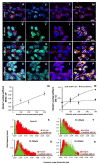
Mitochondrial DNA release and co-localisation analysis of mitochondria (mt) and dsDNA in PA-treated hCMEC/D3s. (AD) Far-left column—Representative images of hCMEC/D3s stained with the dsDNA marker PicoGreen, DAPI (blue) and the mt-specific marker Mito-Tracker Red. (EH) Centre-left column—hCMEC/D3 cells showing tracks with the dsDNA marker PicoGreen and DAPI (blue). (IL) Centre-right column—hCMEC/D3 cells showing tracks labelled with DAPI (blue) and Mito-Tracker Red. (MP) Far-right column—hCMEC/D3 cells showing leakage of dsDNA (depicted by yellow pixels), which increases in the cell’s cytosol with the increasing PA concentration. Open arrowheads (MP) indicate examples where the increase in dsDNA in the cytosol is apparent, and thus co-localisation (yellow area) between dsDNA and mt is evident. (Q) The positive correlation of the increasing co-occurrence between mt and dsDNA, across the PA treatment. (R) Coefficient M1 (dsDNA/mt) indicates that the fraction of dsDNA that overlaps with mt increased with the PA concentration, revealing a clear dose-dependent relationship (n = 6–8). Coefficient M2 (mt/dsDNA) demonstrates that the fraction of mt overlapping with dsDNA did not change across PA concentrations (n = 6–8). (SV) Cytosolic pixel intensity analysis showed a shift of the ‘green’ peak to the right, supporting that dsDNA increased with the PA concentration. Pixel profiles were extracted from where the open arrowheads are indicated (MP). Pixel frequency is normalised between 0 and 1, and mt represents a logged average from across the PA treatments. Scale bar shown on immunofluorescent images represents 10 μm for images (AP).
Figure 7
Figure 7
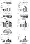
Effect of PA treatment on cGAS-STING signalling and downstream ICAM and IFN-β production in hCMEC/D3s. (A,B) Effect of palmitic acid treatment on cGAS and STING. Dose response effect of PA on (C) TBK-1, (D) pIRF3 and (E) ICAM production, n = 5–6 experiments/condition. (F) Release of IFN-β from cells, n = 5 replicates. Data are expressed as mean% change ± SEM from untreated cells. Mann–Whitney test, * denotes p-values < 0.05. Dots denote individual subject values.
Figure 8
Figure 8
Schematic diagram of activation of the cGAS-STING pathway in the CNS. Cytosolic DNA is recognised by cGAS, leading to formation of a cGAS-DNA complex. This leads to synthesis of a 2′ 3′-cGAMP–STING ligand. Activated STING translocates from the ER to the proximity of the Golgi, where STING is post-translationally modified (palmitoylated and phosphorylated) by TBK-1. This provokes STING to recruit IRF3, leading to IRF3 phosphorylation and dimerization, and the pIRF dimer travels to the nucleus to drive expression of both type I interferons and ICAM expression, as well as NFκ-B inflammatory genes. This diagram was created with BioRender.com.

References

    1. Reinert L.S., Lopusna K., Winther H., Sun C., Thomsen M.K., Nandakumar R., Mogensen T.H., Meyer M., Vaegter C., Nyengaard J.R., et al. Sensing of HSV-1 by the cGAS-STING pathway in microglia orchestrates antiviral defence in the CNS. Nat. Commun. 2016;7:13348. doi: 10.1038/ncomms13348. - DOI - PMC - PubMed
    1. Zierhut C., Funabiki H. Regulation and Consequences of cGAS Activation by Self-DNA. Trends Cell. Biol. 2020;30:594–605. doi: 10.1016/j.tcb.2020.05.006. - DOI - PMC - PubMed
    1. Gutowski N.J., Ferecsko A.S., Smallwood M., Holley J., Newcombe J., Moore A., Eggleton P. Human neurodegenerative CNS tissues show robust expression of cGAS-STING pathway components in association with inflammation. Eur. J. Neurol. 2019;26:717.
    1. Ahmad L., Zhang S.Y., Casanova J.L., Sancho-Shimizu V. Human TBK1: A Gatekeeper of Neuroinflammation. Trends Mol. Med. 2016;22:511–527. doi: 10.1016/j.molmed.2016.04.006. - DOI - PMC - PubMed
    1. Oakes J.A., Davies M.C., Collins M.O. TBK1: A new player in ALS linking autophagy and neuroinflammation. Mol. Brain. 2017;10:5. doi: 10.1186/s13041-017-0287-x. - DOI - PMC - PubMed