Review
. 2023 May 17;11(5):1461.
doi: 10.3390/biomedicines11051461.

Cerebrospinal Fluid-Basic Concepts Review

Affiliations
Review

Cerebrospinal Fluid-Basic Concepts Review

Natalia Czarniak et al. Biomedicines. .

Abstract

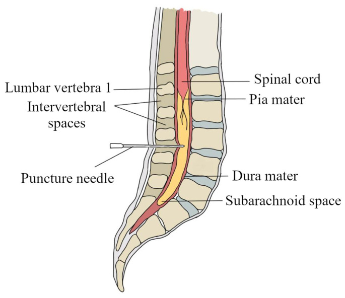
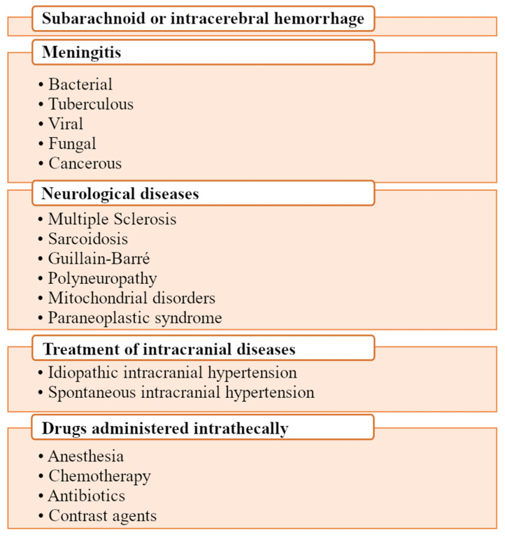
Cerebrospinal fluid plays a crucial role in protecting the central nervous system (CNS) by providing mechanical support, acting as a shock absorber, and transporting nutrients and waste products. It is produced in the ventricles of the brain and circulates through the brain and spinal cord in a continuous flow. In the current review, we presented basic concepts related to cerebrospinal fluid history, cerebrospinal fluid production, circulation, and its main components, the role of the blood-brain barrier and the blood-cerebrospinal fluid barrier in the maintenance of cerebrospinal fluid homeostasis, and the utility of Albumin Quotient (QAlb) evaluation in the diagnosis of CNS diseases. We also discussed the collection of cerebrospinal fluid (type, number of tubes, and volume), time of transport to the laboratory, and storage conditions. Finally, we briefly presented the role of cerebrospinal fluid examination in CNS disease diagnosis of various etiologies and highlighted that research on identifying cerebrospinal fluid biomarkers indicating disease presence or severity, evaluating treatment effectiveness, and enabling understanding of pathogenesis and disease mechanisms is of great importance. Thus, in our opinion, research on cerebrospinal fluid is still necessary for both the improvement of CNS disease management and the discovery of new treatment options.

Keywords: cerebrospinal fluid; cerebrospinal fluid biomarkers; cerebrospinal fluid examination; cerebrospinal fluid storage; cerebrospinal fluid transport conditions; the blood-cerebrospinal fluid barrier; the blood–brain barrier.


Conflict of interest statement

The authors declare no conflict of interest.

Figures

Figure 1
Figure 1
Cerebrospinal fluid production. Its production is based on the active exchange of ions and H2O between the interstitial space of the choroid plexus and the cerebrospinal fluid. Carbonic anhydrase catalyzes the conversion of H2O and CO2 to H+ and HCO3 ions. Ion carrier proteins transport Na+, Cl, and HCO3 ions from the extracellular fluid through the basolateral membrane into the choroid plexus epithelial cells and then, after intracellular circulation, through the apical membrane of the choroid plexus epithelial cells into the cerebrospinal fluid. H2O enters the choroid plexus epithelial cells mainly through AQP1 as a result of the osmotic pressure gradient. AE2—anion exchange protein 2; AQP1—aquaporin 1; Cl—chloride ions; Clir—inward-rectifying chloride channel; CO2—carbon dioxide; CSF—cerebrospinal fluid; H+—hydrogen ions; H2O—hydrogen monoxide, water; HCO3—bicarbonate ions; K+—potassium ions; KCC1—potassium chloride cotransporter 1; KCC4—potassium-chloride co-transporter 4; Kir—inward-rectifier potassium channel; Na+—sodium ions; ATPase Na+/K+—sodium-potassium pump; NBCn1—sodium bicarbonate co-transporter 1; NBCn2—sodium bicarbonate co-transporter 2; NHE—sodium-hydrogen exchanger; NKCC1—sodium potassium chloride co-transporter 1; VRAC—volume-regulated anion channel.
Figure 2
Figure 2
Cerebrospinal fluid flow. Cerebrospinal fluid is mainly produced in the lateral ventricles of the brain, while being produced to a smaller extent in the third and fourth ventricles. From the lateral ventricles of the brain, the cerebrospinal fluid flows through Monroe’s foramen into the third ventricle, and it flows from there through the aqueduct of Sylvius into the fourth ventricle, from where it flows through the Magendi’s foramen and two lateral foramina of Luschka into the subarachnoid space of the brain and the spinal cord. Cerebrospinal fluid is mainly absorbed through the arachnoid granulations into the dural venous sinuses and from there into the blood. Arrows shows direction of cerebrospinal fluid flow.
Figure 3
Figure 3
Examples of substances present in cerebrospinal fluid. BDNF—brain-derived neurotrophic factor; Ca2+—calcium ions; Cl—chloride ions; HCO3—bicarbonate ions; IGF-2—insulin-like growth factor 2; K+—potassium ions; Mg2+—magnesium ions; Mn2+—manganate ions; Na+—sodium ions.
Figure 4
Figure 4
Structural components of the blood–brain barrier. Blue—astrocyte, yellow—pericyte, gray—neuron, red—endothelial cells.
Figure 5
Figure 5
Illustration of the junction between two brain capillary endothelial cells that make up the blood–brain barrier. Tight junctions (TJs) consist of various subunits of transport proteins such as occludins, claudins, cadherins, and JAM adhesion molecules. Adherens junctions (AJs) are responsible for the initiation and stabilization of intercellular adhesions and regulate the actin cytoskeleton of endothelial cells of brain capillaries. ESAM—endothelial cell-selective adhesion molecule; JAM-A—junctional adhesion molecule; PECAM-1—platelet–endothelial cell adhesion molecule-1; VE-cadherin—vascular endothelial-cadherin; ZO—zonula occludens.
Figure 6
Figure 6
A schematic diagram presenting the blood–cerebrospinal fluid barrier (BCB). Functionally, BCB is a set of mechanisms that allow proteins to flow from the blood into the cerebrospinal fluid. BCB is formed by epithelial cells of the choroid plexus of the four ventricles of the brain and epithelial subarachnoid structures directed in the intracranial areas and the spine to the cerebrospinal fluid space.
Figure 7
Figure 7
Diagram showing the site of cerebrospinal fluid collection by lumbar puncture. To perform a lumbar puncture, a puncture needle should be placed between the 3rd and 4th or 4th and 5th lumbar vertebrae. The needle is then inserted into the subarachnoid space where the cerebrospinal fluid is located.
Figure 8
Figure 8
Indications for cerebrospinal fluid collection. The main purpose of collecting cerebrospinal fluid is the diagnosis of CNS diseases. In addition, drugs that do not penetrate from the blood into the CNS can also be administered via a lumbar puncture.

References

    1. Zambito Marsala S., Gioulis M., Pistacchi M. Cerebrospinal fluid and lumbar puncture: The story of a necessary procedure in the history of medicine. Neurol. Sci. 2015;36:1011–1015. doi: 10.1007/s10072-015-2104-6. - DOI - PubMed
    1. Liddelow S.A. Development of the choroid plexus and blood-CSF barrier. Front. Neurosci. 2015;9:32. doi: 10.3389/fnins.2015.00032. - DOI - PMC - PubMed
    1. Van Middendorp J.J., Sanchez G.M., Burridge A.L. The Edwin Smith papyrus: A clinical reappraisal of the oldest known document on spinal injuries. Eur. Spine J. 2010;19:1815–1823. doi: 10.1007/s00586-010-1523-6. - DOI - PMC - PubMed
    1. Hajdu S.I. A note from history: Discovery of the cerebrospinal fluid. Ann. Clin. Lab. Sci. 2003;33:334–336. - PubMed
    1. Herbowski L. The Maze of the Cerebrospinal Fluid Discovery. Anat. Res. Int. 2013;2013:596027. doi: 10.1155/2013/596027. - DOI - PMC - PubMed

LinkOut - more resources