. 2023 May 17;11(5):1464.
doi: 10.3390/biomedicines11051464.

Neurophysiological Markers of Premotor-Motor Network Plasticity Predict Motor Performance in Young and Older Adults

Affiliations

Neurophysiological Markers of Premotor-Motor Network Plasticity Predict Motor Performance in Young and Older Adults

Sonia Turrini et al. Biomedicines. .

Abstract

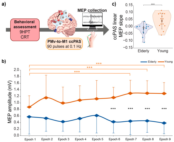
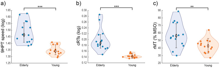
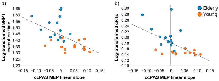
Aging is commonly associated with a decline in motor control and neural plasticity. Tuning cortico-cortical interactions between premotor and motor areas is essential for controlling fine manual movements. However, whether plasticity in premotor-motor circuits predicts hand motor abilities in young and elderly humans remains unclear. Here, we administered transcranial magnetic stimulation (TMS) over the ventral premotor cortex (PMv) and primary motor cortex (M1) using the cortico-cortical paired-associative stimulation (ccPAS) protocol to manipulate the strength of PMv-to-M1 connectivity in 14 young and 14 elderly healthy adults. We assessed changes in motor-evoked potentials (MEPs) during ccPAS as an index of PMv-M1 network plasticity. We tested whether the magnitude of MEP changes might predict interindividual differences in performance in two motor tasks that rely on premotor-motor circuits, i.e., the nine-hole pegboard test and a choice reaction task. Results show lower motor performance and decreased PMv-M1 network plasticity in elderly adults. Critically, the slope of MEP changes during ccPAS accurately predicted performance at the two tasks across age groups, with larger slopes (i.e., MEP increase) predicting better motor performance at baseline in both young and elderly participants. These findings suggest that physiological indices of PMv-M1 plasticity could provide a neurophysiological marker of fine motor control across age-groups.

Keywords: aging; connectivity; motor cortex; motor performance; plasticity; premotor cortex; transcranial magnetic stimulation.


Conflict of interest statement

The authors declare no conflict of interest.

Figures

Figure 1
Figure 1
(a) Experimental design. Behavioral assessment was followed by the administration of a ccPAS protocol over the left PMv and M1. For each paired PMv-M1 stimulation of the ccPAS protocol, an MEP was collected from the right FDI; (b) mean MEP amplitudes recorded during the ccPAS in elderly (blue) and young (orange) participants along 9 epochs; (c) linear slope of MEP increase during ccPAS in the two groups. Error bars represent standard deviations; *** p ≤ 0.001.
Figure 2
Figure 2
(a) 9HPT performance; (b) cRTs; and (c) rMT in young (orange) and elderly (blue) individuals. Error bars represent standard deviations; ** p ≤ 0.01; *** p ≤ 0.001.
Figure 3
Figure 3
Relation between the neurophysiological marker of STDP (ccPAS MEP linear slope) and motor performance assessed at baseline. The STDP index predicts both 9HPT execution times (a) and cRTs (b) execution times across age groups, showing that larger MEP slope (reflecting greater plasticity) is associated with faster motor performance at baseline. Orange dots represent young participants (N = 14) and blue dots represent elderly participants (N = 14). Dashed lines depict the regression line of the significant predictor ccPAS MEP linear slope on 9HPT execution times (a) and cRTs (b) across groups.

References

    1. Turrini S., Wong B., Eldaief M., Press D.Z., Sinclair D.A., Koch G., Avenanti A., Santarnecchi E. The Multifactorial Nature of Healthy Brain Ageing: Brain Changes, Functional Decline and Protective Factors. Ageing Res. Rev. 2023;88:101939. doi: 10.1016/j.arr.2023.101939. - DOI - PubMed
    1. Damoiseaux J.S. Effects of Aging on Functional and Structural Brain Connectivity. Neuroimage. 2017;160:32–40. doi: 10.1016/j.neuroimage.2017.01.077. - DOI - PubMed
    1. Good C.D., Johnsrude I.S., Ashburner J., Henson R.N.A., Friston K.J., Frackowiak R.S.J. A Voxel-Based Morphometric Study of Ageing in 465 Normal Adult Human Brains. NeuroImage. 2001;14:21–36. doi: 10.1006/nimg.2001.0786. - DOI - PubMed
    1. Salat D.H., Buckner R.L., Snyder A.Z., Greve D.N., Desikan R.S.R., Busa E., Morris J.C., Dale A.M., Fischl B. Thinning of the Cerebral Cortex in Aging. Cereb. Cortex. 2004;14:721–730. doi: 10.1093/cercor/bhh032. - DOI - PubMed
    1. Resnick S.M., Pham D.L., Kraut M.A., Zonderman A.B., Davatzikos C. Longitudinal Magnetic Resonance Imaging Studies of Older Adults: A Shrinking Brain. J. Neurosci. 2003;23:3295–3301. doi: 10.1523/JNEUROSCI.23-08-03295.2003. - DOI - PMC - PubMed

LinkOut - more resources