. 2023 May 19;11(5):1481.
doi: 10.3390/biomedicines11051481.

Cognitive and Cellular Effects of Combined Organophosphate Toxicity and Mild Traumatic Brain Injury

Affiliations

Cognitive and Cellular Effects of Combined Organophosphate Toxicity and Mild Traumatic Brain Injury

Dor Freidin et al. Biomedicines. .

Abstract

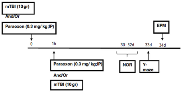
Traumatic brain injury (TBI) is considered the most common neurological disorder among people under the age of 50. In modern combat zones, a combination of TBI and organophosphates (OP) can cause both fatal and long-term effects on the brain. We utilized a mouse closed-head TBI model induced by a weight drop device, along with OP exposure to paraoxon. Spatial and visual memory as well as neuron loss and reactive astrocytosis were measured 30 days after exposure to mild TBI (mTBI) and/or paraoxon. Molecular and cellular changes were assessed in the temporal cortex and hippocampus. Cognitive and behavioral deficits were most pronounced in animals that received a combination of paraoxon exposure and mTBI, suggesting an additive effect of the insults. Neuron survival was reduced in proximity to the injury site after exposure to paraoxon with or without mTBI, whereas in the dentate gyrus hilus, cell survival was only reduced in mice exposed to paraoxon prior to sustaining a mTBI. Neuroinflammation was increased in the dentate gyrus in all groups exposed to mTBI and/or to paraoxon. Astrocyte morphology was significantly changed in mice exposed to paraoxon prior to sustaining an mTBI. These results provide further support for assumptions concerning the effects of OP exposure following the Gulf War. This study reveals additional insights into the potentially additive effects of OP exposure and mTBI, which may result in more severe brain damage on the modern battlefield.

Keywords: cognitive and behavioral tests; mTBI; neuroinflammation; neuronal loss; organophosphates.


Conflict of interest statement

The authors declare no conflict of interest.

Figures

Figure 1
Figure 1
Timeline.
Figure 2
Figure 2
Behavioral test scores. (A) NOR test: differences in visual recognition memory performance between mice in the control (n = 12), mTBI (n = 12), paraoxon (n = 11), paraoxon + mTBI (n = 10), and mTBI + paraoxon (n = 10) groups. (B) Y-maze test: differences in spatial memory performance between mice in the control (n = 12), mTBI (n = 12), paraoxon (n = 11), paraoxon + mTBI (n = 10), and mTBI + paraoxon (n = 10) groups. * p < 0.05, ** p < 0.01, *** p < 0.001.
Figure 3
Figure 3
NeuN+ counts in the cortex and hippocampus of control (n = 5), mTBI (n = 5), paraoxon (n = 5), paraoxon + mTBI (n = 5), and mTBI + paraoxon (n = 5) mice. Paraoxon exposure before or after mTBI led to a significant decrease in the density of NeuN+ neurons compared to control and mTBI tissues in the cortex. (A) Quantification of total surface area labeled with NeuN in the temporal cortex. (B) Quantification of total surface area labeled with NeuN in the DGH. (C) Representative images of immunohistochemical staining in the temporal cortex (upper panel) and DGH (lower panel). NeuN+ cells are shown in red, and nuclei are shown in blue. Yellow lines outline the hilus region of the DG. * p < 0.05, ** p < 0.01, *** p < 0.001.
Figure 4
Figure 4
Astrocyte changes in the cortex and hippocampus of control (n = 5), mTBI (n = 5), paraoxon (n = 5), paraoxon + mTBI (n = 5), and mTBI + paraoxon (n = 5) mice. Paraoxon increases active astrocyte expression in the DGH only and changes astrocyte morphology before mTBI. The graphs present quantifications of: (A) GFAP intensity in the temporal cortex; (B) GFAP intensity in the DGH; (C) astrocyte counts in the temporal cortex; (D) astrocyte counts in the DGH; (E) astrocyte morphology in the temporal cortex; and (F) astrocyte morphology in the DG. Representative images of immunohistochemical staining in the DGH and the temporal cortex are presented in (G). GFAP-positive cells are shown in green, and nuclei are shown in blue. Scale bars are 25 μm. * p < 0.05, ** p < 0.01.

References

    1. Holm L., Cassidy J.D., Carroll L.J., Borg J. Summary of the WHO Collaborating Centre for Neurotrauma Task Force on Mild Traumatic Brain Injury. J. Rehabil. Med. 2005;37:137–141. doi: 10.1080/16501970510027321. - DOI - PubMed
    1. Parikh S., Koch M., Narayan R.K. Traumatic brain injury. Int. Anesthesiol. Clin. 2007;45:119–135. doi: 10.1097/AIA.0b013e318078cfe7. - DOI - PubMed
    1. Greve M.W., Zink B.J. Pathophysiology of traumatic brain injury. Mt. Sinai. J. Med. 2009;76:97–104. doi: 10.1002/msj.20104. - DOI - PubMed
    1. Lucke-Wold B.P., Logsdon A.F., Nguyen L., Eltanahay A., Turner R.C., Bonasso P., Knotts C., Moeck A., Maroon J.C., Bailes J.E. Supplements, nutrition, and alternative therapies for the treatment of traumatic brain injury. Nutr. Neurosci. 2018;21:79–91. doi: 10.1080/1028415X.2016.1236174. - DOI - PMC - PubMed
    1. Moppett I.K. Traumatic brain injury: Assessment, resuscitation and early management. BJA Br. J. Anaesth. 2007;99:18–31. doi: 10.1093/bja/aem128. - DOI - PubMed

LinkOut - more resources