

Detailed Courses and Pathological Findings of Colonic Perforation without Diverticula in Sisters with Musculocontractural Ehlers-Danlos Syndrome Caused by Pathogenic Variant in CHST14 (mcEDS- CHST14)
- PMID: 37239439
- PMCID: PMC10218258
- DOI: 10.3390/genes14051079
Detailed Courses and Pathological Findings of Colonic Perforation without Diverticula in Sisters with Musculocontractural Ehlers-Danlos Syndrome Caused by Pathogenic Variant in CHST14 (mcEDS- CHST14)
Abstract
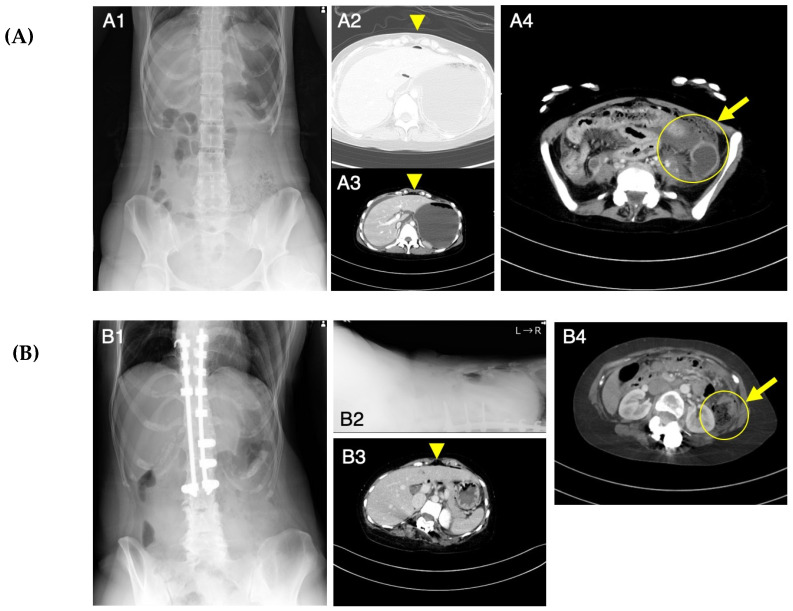
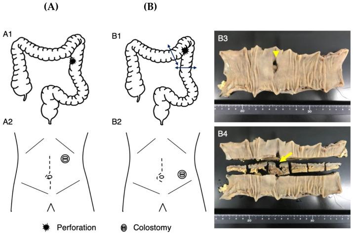
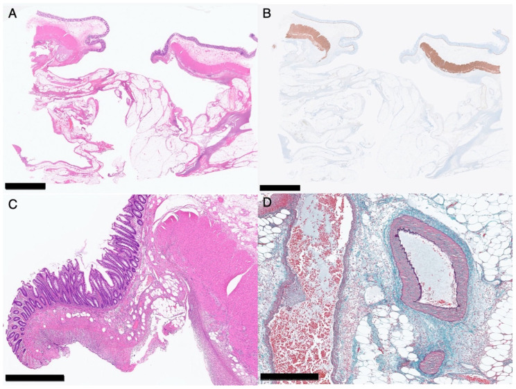
Musculocontractural Ehlers-Danlos syndrome (mcEDS) is a heritable connective tissue disorder characterized by multiple congenital malformations and progressive connective-tissue-fragility-related manifestations in the cutaneous, skeletal, cardiovascular, visceral, ocular, and gastrointestinal systems. It is caused by pathogenic variants in the carbohydrate sulfotransferase 14 gene (mcEDS-CHST14) or in the dermatan sulfate epimerase gene (mcEDS-DSE). As gastrointestinal complications of mcEDS-CHST14, diverticula in the colon, small intestine, or stomach have been reported, which may lead to gastrointestinal perforation, here, we describe sisters with mcEDS-CHST14, who developed colonic perforation with no evidence of diverticula and were successfully treated through surgery (a resection of perforation site and colostomy) and careful postoperative care. A pathological investigation did not show specific abnormalities of the colon at the perforation site. Patients with mcEDS-CHST14 aged from the teens to the 30s should undergo not only abdominal X-ray photography but also abdominal computed tomography when they experience abdominal pain.
Keywords: carbohydrate sulfotransferase 14 (CHST14); diverticulum; gastrointestinal perforation; musculocontractural Ehlers–Danlos syndrome (mcEDS).
Conflict of interest statement
The authors declare no conflict of interest. T.K. (Tomoki Kosho) is a member of an endowed chair named “Division of Clinical Sequencing, Shinshu University School of Medicine” sponsored by BML Inc. and Life Technologies Japan Ltd. of Thermo Fisher Scientific Inc.
Figures
Similar articles
-
Recent Advances in the Pathophysiology of Musculocontractural Ehlers-Danlos Syndrome.Genes (Basel). 2019 Dec 29;11(1):43. doi: 10.3390/genes11010043. Genes (Basel). 2019. PMID: 31905796 Free PMC article. Review.
-
Mouse Models of Musculocontractural Ehlers-Danlos Syndrome.Genes (Basel). 2023 Feb 8;14(2):436. doi: 10.3390/genes14020436. Genes (Basel). 2023. PMID: 36833362 Free PMC article. Review.
-
Collagen Network Formation in In Vitro Models of Musculocontractural Ehlers-Danlos Syndrome.Genes (Basel). 2023 Jan 24;14(2):308. doi: 10.3390/genes14020308. Genes (Basel). 2023. PMID: 36833235 Free PMC article.
-
Delineation of musculocontractural Ehlers-Danlos Syndrome caused by dermatan sulfate epimerase deficiency.Mol Genet Genomic Med. 2020 May;8(5):e1197. doi: 10.1002/mgg3.1197. Epub 2020 Mar 4. Mol Genet Genomic Med. 2020. PMID: 32130795 Free PMC article.
-
Clinical and molecular features of 66 patients with musculocontractural Ehlers-Danlos syndrome caused by pathogenic variants in CHST14 (mcEDS-CHST14).J Med Genet. 2022 Sep;59(9):865-877. doi: 10.1136/jmedgenet-2020-107623. Epub 2021 Nov 23. J Med Genet. 2022. PMID: 34815299 Free PMC article.
Cited by
-
Perforation of the Sigmoid Colon due to Diverticular Rupture in a Woman With FKBP14-Related Kyphoscoliotic Ehlers-Danlos Syndrome: A Case Report.Clin Case Rep. 2025 May 6;13(5):e70480. doi: 10.1002/ccr3.70480. eCollection 2025 May. Clin Case Rep. 2025. PMID: 40330264 Free PMC article.
-
Surgical management of endometrial cancer in patient with musculocontractural Ehlers-Danlos Syndrome harboring pathogenic variants in CHST14 (mcEDS-CHST14): A case report.Gynecol Oncol Rep. 2024 Dec 31;57:101675. doi: 10.1016/j.gore.2024.101675. eCollection 2025 Feb. Gynecol Oncol Rep. 2024. PMID: 39867552 Free PMC article.
-
Carbohydrate sulfotransferase 14 gene deletion induces dermatan sulfate deficiency and affects collagen structure and bowel contraction.PLoS One. 2025 May 6;20(5):e0320943. doi: 10.1371/journal.pone.0320943. eCollection 2025. PLoS One. 2025. PMID: 40327642 Free PMC article.
References
-
- Minatogawa M., Unzaki A., Morisaki H., Syx D., Sonoda T., Janecke A.R., Slavotinek A., Voermans N.C., Lacassie Y., Mendoza-Londono R., et al. Clinical and Molecular Features of 66 Patients with Musculocontractural Ehlers-Danlos Syndrome Caused by Pathogenic Variants in CHST14 (mcEDS-CHST14) J. Med. Genet. 2022;59:865–877. doi: 10.1136/jmedgenet-2020-107623. - DOI - PMC - PubMed
Publication types
MeSH terms
Substances
LinkOut - more resources
Full Text Sources
Medical
Research Materials
