. 2023 May 12;24(10):8648.
doi: 10.3390/ijms24108648.

Study on the Mechanism of MC5R Participating in Energy Metabolism of Goose Liver

Affiliations

Study on the Mechanism of MC5R Participating in Energy Metabolism of Goose Liver

Jinqi Zhang et al. Int J Mol Sci. .

Abstract

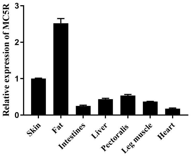
Nutrition and energy levels have an important impact on animal growth, production performance, disease occurrence and health recovery. Previous studies indicate that melanocortin 5 receptor (MC5R) is mainly involved in the regulations of exocrine gland function, lipid metabolism and immune response in animals. However, it is not clear how MC5R participates in the nutrition and energy metabolism of animals. To address this, the widely used animal models, including the overfeeding model and the fasting/refeeding model, could provide an effective tool. In this study, the expression of MC5R in goose liver was first determined in these models. Goose primary hepatocytes were then treated with nutrition/energy metabolism-related factors (glucose, oleic acid and thyroxine), which is followed by determination of MC5R gene expression. Moreover, MC5R was overexpressed in goose primary hepatocytes, followed by identification of differentially expressed genes (DEGs) and pathways subjected to MC5R regulation by transcriptome analysis. At last, some of the genes potentially regulated by MC5R were also identified in the in vivo and in vitro models, and were used to predict possible regulatory networks with PPI (protein-protein interaction networks) program. The data showed that both overfeeding and refeeding inhibited the expression of MC5R in goose liver, while fasting induced the expression of MC5R. Glucose and oleic acid could induce the expression of MC5R in goose primary hepatocytes, whereas thyroxine could inhibit it. The overexpression of MC5R significantly affected the expression of 1381 genes, and the pathways enriched with the DEGs mainly include oxidative phosphorylation, focal adhesion, ECM-receptor interaction, glutathione metabolism and MAPK signaling pathway. Interestingly, some pathways are related to glycolipid metabolism, including oxidative phosphorylation, pyruvate metabolism, citrate cycle, etc. Using the in vivo and in vitro models, it was demonstrated that the expression of some DEGs, including ACSL1, PSPH, HMGCS1, CPT1A, PACSIN2, IGFBP3, NMRK1, GYS2, ECI2, NDRG1, CDK9, FBXO25, SLC25A25, USP25 and AHCY, was associated with the expression of MC5R, suggesting these genes may mediate the biological role of MC5R in these models. In addition, PPI analysis suggests that the selected downstream genes, including GYS2, ECI2, PSPH, CPT1A, ACSL1, HMGCS1, USP25 and NDRG1, participate in the protein-protein interaction network regulated by MC5R. In conclusion, MC5R may mediate the biological effects caused by changes in nutrition and energy levels in goose hepatocytes through multiple pathways, including glycolipid-metabolism-related pathways.

Keywords: MC5R; differentially expressed gene; energy metabolism; goose; liver.


Conflict of interest statement

The authors declare that this study was carried out without any commercial or financial relationships that could be construed as a potential conflict of interest.

Figures

Figure 1
Figure 1
The mRNA expression level of MC5R gene in different embryonic tissues of Landes geese. Note: The mRNA expression level was determined by RT-qPCR and presented as fold change over skin tissue. The internal reference gene was glyceraldehyde-3-phosphate dehydrogenase (GAPDH). The embryos were from the fertilized eggs cultured for 23 days. n = 3.
Figure 2
Figure 2
The mRNA and protein expression levels of MC5R in the livers of the overfed versus normally fed Landes geese on the 12th (12D) and 24th days (24D) of overfeeding. Note: (A) The mRNA expression level was determined by RT-qPCR and presented as fold change over the control group (the normally fed geese). The internal reference gene was GAPDH. n = 6. (B) The protein expression level was determined by immunoblot assay. The internal reference gene was α-tubulin. The protein samples were from the livers of geese on the 24th day of overfeeding. (C) Quantification of the immunoblots. *, *** indicate p < 0.05 and 0.001, respectively.
Figure 3
Figure 3
The mRNA expression level of MC5R gene in the livers of the fasted and refed versus control Landes geese. Note: The mRNA expression level was determined by RT-qPCR and presented as fold change over the control group. The internal reference gene was GAPDH. n = 8. *, **, *** indicate p < 0.05, 0.01 and 0.001, respectively.
Figure 4
Figure 4
The mRNA expression level of MC5R in the treated versus control primary hepatocytes of goose. Note: The factors that are related to nutrition or energy metabolism include (A) glucose, (B) sodium oleate and (C) sodium thyroxine. The mRNA expression level was determined by RT-qPCR and presented as fold change over the control group. The internal reference gene was GAPDH. n = 6. *, ** indicate p < 0.05 and 0.01, respectively. ‘ns’ denotes p > 0.05.
Figure 5
Figure 5
Overexpression of MC5R gene in goose primary hepatocytes. Note: The (A) mRNA and (B) protein expression levels of MC5R in goose primary hepatocytes treated with MC5R overexpression vector vs. empty vector (the control group). (C) Quantification of the immunoblots. The mRNA expression level was determined by RT-qPCR and presented as fold change over the control group. The internal reference gene was ubiquitin C (UBC). n = 6. The protein expression level was determined by immunoblot assay. The internal reference gene was GAPDH. *, *** indicate p < 0.05 and 0.001, respectively.
Figure 6
Figure 6
The dot chart shows the results from KEGG enrichment analysis. Note: (A) The results of KEGG enrichment analysis with the upregulated DEGs. (B) The results of KEGG enrichment analysis with the downregulated DEGs. The abscissa indicates the ratio of the number of the annotated DEGs in a specified KEGG pathway to the total number of annotated DEGs, and the ordinate indicates the KEGG pathways. The colors denote the adjusted p-value (p-adj), and the sizes of the dots denote the number of DEGs. The DEGs were identified in goose primary hepatocytes transfected with MC5R overexpression vectors vs. empty vectors (n = 4).
Figure 6
Figure 6
The dot chart shows the results from KEGG enrichment analysis. Note: (A) The results of KEGG enrichment analysis with the upregulated DEGs. (B) The results of KEGG enrichment analysis with the downregulated DEGs. The abscissa indicates the ratio of the number of the annotated DEGs in a specified KEGG pathway to the total number of annotated DEGs, and the ordinate indicates the KEGG pathways. The colors denote the adjusted p-value (p-adj), and the sizes of the dots denote the number of DEGs. The DEGs were identified in goose primary hepatocytes transfected with MC5R overexpression vectors vs. empty vectors (n = 4).
Figure 7
Figure 7
The mRNA expression level of some specifically selected DEGs in goose primary hepatocytes transfected with MC5R overexpression vector vs. empty vector. Note: The mRNA expression level was determined by RT-qPCR and presented as fold change over the control group. The internal reference gene was UBC. n = 6. *,**, *** indicate p < 0.05, 0.01 and 0.001, respectively.
Figure 8
Figure 8
The mRNA expression level of MC5R (A) and some specifically selected DEGs (B) in goose primary hepatocytes transfected with siRNA targeting MC5R (siMC5R) versus the control negative siRNA (control). Note: The mRNA expression level was determined by RT-qPCR and presented as fold change over the control group. The internal reference gene was UBC. n = 6. *,**, *** indicate p < 0.05, 0.01 and 0.001, respectively. ‘ns’ denotes p > 0.05.
Figure 9
Figure 9
The mRNA expression level of some specifically selected DEGs in goose primary hepatocytes treated with 100 mM/L glucose versus the control group (0 mM/L glucose). Note: The mRNA expression level was determined by RT-qPCR and presented as fold change over the control group. The internal reference gene was GAPDH. n = 6. *,**, *** indicate p < 0.05, 0.01 and 0.001, respectively. ‘ns’ denotes p > 0.05.
Figure 10
Figure 10
The mRNA expression level of some specifically selected DEGs in the livers of the overfed versus normally fed geese on the 24th day of overfeeding. Note: The mRNA expression level was determined by RT-qPCR and presented as fold change over the control group (the normally fed geese). The internal reference gene was GAPDH. n = 6. *,**, *** indicate p < 0.05, 0.01 and 0.001, respectively. ‘ns’ denotes p > 0.05.
Figure 11
Figure 11
The mRNA expression level of some specifically selected DEGs in the livers of the refed versus fasted geese. Note: The mRNA expression level was determined by RT-qPCR and presented as fold change over the fasted group. The internal reference gene was GAPDH. n = 8. *,**, *** indicate p < 0.05, 0.01 and 0.001, respectively.
Figure 12
Figure 12
The chart shows the protein–protein interaction network regulated by MC5R based on PPI analysis. Note: Colors ranging from yellow to red indicate an increase in degree of connectivity, and a larger degree indicates a greater connectivity to other genes.

References

    1. Trefts E., Gannon M., Wasserman D.H. The liver. Curr. Biol. 2017;27:R1147–R1151. doi: 10.1016/j.cub.2017.09.019. - DOI - PMC - PubMed
    1. Jones J.G. Hepatic glucose and lipid metabolism. Diabetologia. 2016;59:1098–1103. doi: 10.1007/s00125-016-3940-5. - DOI - PubMed
    1. Adeva-Andany M.M., Pérez-Felpete N., Fernández-Fernández C., Donapetry-García C., Pazos-García C. Liver glucose metabolism in humans. Biosci. Rep. 2016;36:e00416. doi: 10.1042/BSR20160385. - DOI - PMC - PubMed
    1. Han H.S., Kang G., Kim J.S., Choi B.H., Koo S.H. Regulation of glucose metabolism from a liver-centric perspective. Exp. Mol. Med. 2016;48:e218. doi: 10.1038/emm.2015.122. - DOI - PMC - PubMed
    1. Zeng Q.F., Cherry P., Doster A., Murdoch R., Adeola O., Applegate T.J. Effect of dietary energy and protein content on growth and carcass traits of Pekin ducks. Poult. Sci. 2015;94:384–394. doi: 10.3382/ps/peu069. - DOI - PMC - PubMed

LinkOut - more resources