

Identification of Endometrial Cancer-Specific microRNA Biomarkers in Endometrial Fluid
- PMID: 37240034
- PMCID: PMC10218319
- DOI: 10.3390/ijms24108683
Identification of Endometrial Cancer-Specific microRNA Biomarkers in Endometrial Fluid
Abstract
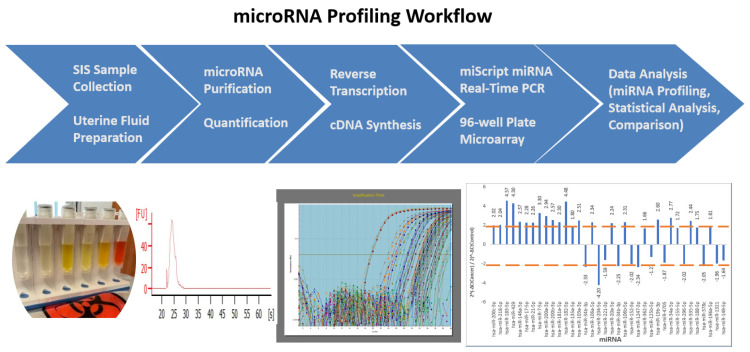
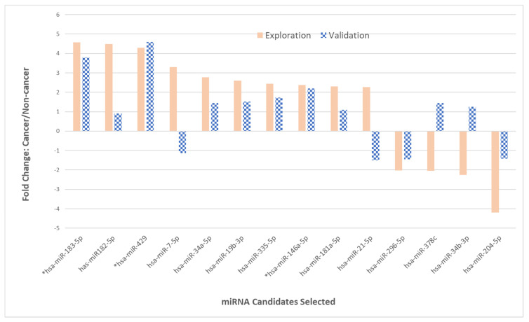
Abnormal uterine bleeding is a common benign gynecological complaint and is also the most common symptom of endometrial cancer (EC). Although many microRNAs have been reported in endometrial carcinoma, most of them were identified from tumor tissues obtained at surgery or from cell lines cultured in laboratories. The objective of this study was to develop a method to detect EC-specific microRNA biomarkers from liquid biopsy samples to improve the early diagnosis of EC in women. Endometrial fluid samples were collected during patient-scheduled in-office visits or in the operating room prior to surgery using the same technique performed for saline infusion sonohysterography (SIS). The total RNA was extracted from the endometrial fluid specimens, followed by quantification, reverse transcription, and real-time PCR arrays. The study was conducted in two phases: exploratory phase I and validation phase II. In total, endometrial fluid samples from 82 patients were collected and processed, with 60 matched non-cancer versus endometrial carcinoma patients used in phase I and 22 in phase II. The 14 microRNA biomarkers, out of 84 miRNA candidates, with the greatest variation in expression from phase I, were selected to enter phase II validation and statistical analysis. Among them, three microRNAs had a consistent and substantial fold-change in upregulation (miR-429, miR-183-5p, and miR-146a-5p). Furthermore, four miRNAs (miR-378c, miR-4705, miR-1321, and miR-362-3p) were uniquely detected. This research elucidated the feasibility of the collection, quantification, and detection of miRNA from endometrial fluid with a minimally invasive procedure performed during a patient in-office visit. The screening of a larger set of clinical samples was necessary to validate these early detection biomarkers for endometrial cancer.
Keywords: downregulation; endometrial biopsy; endometrial cancer (EC); endometrial fluid; exploration; liquid biopsy; microRNA; saline infusion sonohysterography (SIS); upregulation; validation.
Conflict of interest statement
The authors declare no conflict of interest.
Figures


References
-
- Key Statistics for Endometrial Cancer. [(accessed on 10 November 2022)]. Available online: https://www.cancer.org/cancer/endometrial-cancer/about/key-statistics.html.
-
- Simon C., Crawford S.C., Davis J.A., Siddiqui N.A., Caestecker L.D., Gillis C.R., Hole D., Penney G. The waiting time paradox: Population based retrospective study of treatment delay and survival of women with endometrial cancer in Scotland. BMJ. 2002;325:196. doi: 10.1136/bmj.325.7357.196. - DOI - PMC - PubMed
MeSH terms
Substances
LinkOut - more resources
Full Text Sources
