. 2023 May 15;24(10):8806.
doi: 10.3390/ijms24108806.

Asymmetric Dimethylation of Ribosomal S6 Kinase 2 Regulates Its Cellular Localisation and Pro-Survival Function

Affiliations

Asymmetric Dimethylation of Ribosomal S6 Kinase 2 Regulates Its Cellular Localisation and Pro-Survival Function

Mahmoud I Khalil et al. Int J Mol Sci. .

Abstract

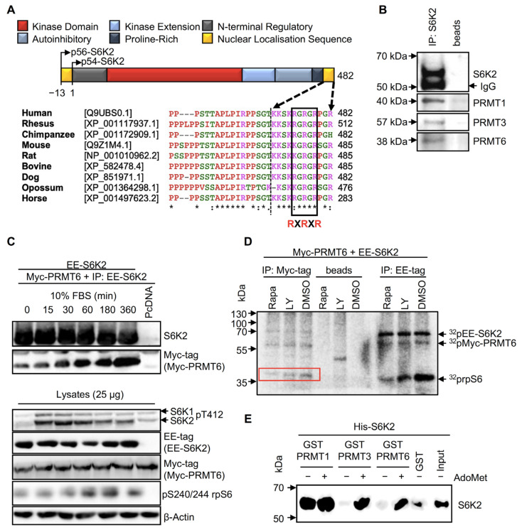
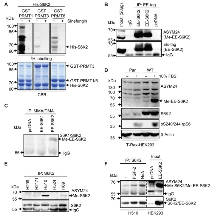
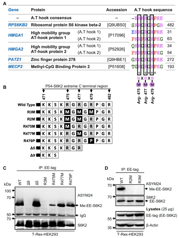
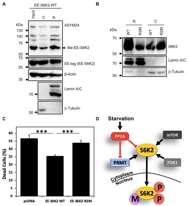
Ribosomal S6 kinases (S6Ks) are critical regulators of cell growth, homeostasis, and survival, with dysregulation of these kinases found to be associated with various malignancies. While S6K1 has been extensively studied, S6K2 has been neglected despite its clear involvement in cancer progression. Protein arginine methylation is a widespread post-translational modification regulating many biological processes in mammalian cells. Here, we report that p54-S6K2 is asymmetrically dimethylated at Arg-475 and Arg-477, two residues conserved amongst mammalian S6K2s and several AT-hook-containing proteins. We demonstrate that this methylation event results from the association of S6K2 with the methyltransferases PRMT1, PRMT3, and PRMT6 in vitro and in vivo and leads to nuclear the localisation of S6K2 that is essential to the pro-survival effects of this kinase to starvation-induced cell death. Taken together, our findings highlight a novel post-translational modification regulating the function of p54-S6K2 that may be particularly relevant to cancer progression where general Arg-methylation is often elevated.

Keywords: AT-hook; SCLC; arginine methylation; methyltransferases; serine/threonine kinases.


Conflict of interest statement

The authors declare no conflict of interest.

Figures

Figure 1
Figure 1
S6K2 interacts with PRMT1, 3, and 6 in vivo and in vitro. (A) Protein domain organisation of S6K2 and alignment of the amino acid sequences in its extreme C-terminus. The conserved Arginine-X-Arginine motif (RXRXR) is boxed, and “X” represents any amino acid. An “*” represents positions which have a single, fully conserved residue. (B) Endogenous S6K2 interacts with PRMTs (1, 3, and 6) in cells. S6K2 was immunoprecipitated from HEK293 and interacting proteins were detected by immunoblotting. Protein A-Sepharose beads (beads) alone were used as a control. (C) The interaction between S6K2 and PRMT6 is induced by serum stimulation. HEK293 cells expressing Myc-PRMT6 and EE-S6K2 or pcDNA3.1 empty vector (pcDNA) were incubated in serum-free media for 24 h prior to treatment with 10% FBS for the indicated times. The lysates were analysed directly or immunoprecipitated with an anti-EE-tag antibody prior to SDS-PAGE/immunoblotting with the indicated primary antibodies. (D) S6K2 activity is immunoprecipitated with PRMT6. Lysates from HEK293 cells expressing Myc-PRMT6 and EE-S6K2 were immunoprecipitated with either anti-EE-tag or anti-Myc-tag antibodies following treatment for 30 min with 100 nM Rapamycin (Rapa), 50 µM LY294002 (Ly), or DMSO (vehicle). The immune complexes were subjected to in vitro kinase assay using 80S ribosomes as substrate in the presence of [γ-32P]ATP prior to SDS-PAGE and autoradiography. The phosphorylated ribosomal S6 proteins (rpS6) were phosphorylated with the immunoprecipitated Myc-PRMT6 (red-boxed). Control IP was achieved by incubating the lysates with protein A-Sepharose beads (beads). (E) PRMT1, PRMT3, and PRMT6 interact with S6K2 in vitro. GST pull-down assays were performed using His-S6K2 with GST, GST-PRMT1, GST-PRMT3, or GST-PRMT6 in the presence or absence of 200 μM AdoMet, followed by immunoblotting with anti-S6K2 antibody. The input is 0.5 μg His-S6K2. (BE) All data shown are representative of a minimum of n = 3 biological repeats.
Figure 2
Figure 2
S6K2 is methylated in vitro and in cells. (A) In vitro methylation of recombinant S6K2. In vitro methylation assays with His-S6K2 and GST-PRMTs (1, 3, or 6) in the presence of [3H]AdoMet, with or without Sinefungin (500 μM). Total amounts of His-S6K2 and GST-PRMTs are shown by Coomassie staining (CBB). (B) Transiently overexpressed S6K2 is methylated in HEK293 cells. Lysates from HEK293 cells expressing EE-S6K2 or pcDNA3.1 empty vector (pcDNA) were immunoprecipitated with an anti-ASYM24 or anti-EE-tag antibody. IgG was used as an immunoprecipitation control. Input: total lysates from HEK293 cells overexpressing EE-S6K2. (C) Methylated EE-S6K2 is immunoprecipitated with an MMA/DMA antibody in T-Rex-HEK293 cells. Lysates from cells overexpressing EE-S6K1, EE-S6K2, or the pcDNA3.1 empty vector (pcDNA) were immunoprecipitated with an anti-Mono/DiMethyl Arginine (MMA/DMA) antibody, followed by immunoblotting with an anti-S6K1 and anti-S6K2 mixture of primary antibodies. (D) S6K2 methylation is induced by serum stimulation. T-Rex-HEK293 cells overexpressing pcDNA3.1 empty vector (Par) or EE-S6K2 (WT) were induced with 1 µg/mL tetracycline for 24 h. The cells were starved in serum-free media (−) for 24 h or stimulated with 10% FBS for 1 h (+) following starvation. The lysates were analysed by SDS-PAGE and immunoblotted with the indicated primary antibodies. (E) S6K2 is methylated in SCLC cell lines. Lysates from the indicated SCLC cell lines were subjected to immunoprecipitation with an anti-S6K2 antibody, followed by immunoblotting with an anti-ASYM24 or anti-S6K2 antibody. (F) S6K2 methylation in H510 cells is induced by FGF2 and sodium arsenite. The lysates from untreated (−), 0.1 ng/mL FGF-2-treated (5 min), or 1 mM sodium arsenite (NaA)-treated (30 min) H510 cells were subjected to immunoprecipitation with an anti-S6K2 antibody, followed by immunoblotting with an anti-ASYM24 or anti-S6K2 antibody. Input: total lysates from HEK293 cells overexpressing EE-S6K2 or the pcDNA3.1 empty vector (pcDNA). (AF) All data shown are representative of n = 3 biological repeats.
Figure 3
Figure 3
S6K2 is methylated at Arg-475 and Arg-477. (A) Sequence alignment of the extreme C-terminal region of S6K2 and selected AT-hook-containing proteins. The putative arginine methylation sites are boxed. RXRXR; consensus sequence for PRMTs substrates. “X” is any amino acid. An “*” represents positions which have a single, fully conserved residue. (B) Schematic representation of S6K2 mutants. Single, double, or triple substitution mutants (Arg to Met or Pro) are generated in addition to two truncated mutants (Δ5 and Δ9). (C,D) S6K2 is dimethylated at Arg-475/477. Lysates from T-Rex-HEK293 cells stably expressing EE-S6K2 wild type (WT), or different mutants were subjected to immunoprecipitation with an anti-EE-tag antibody followed by immunoblotting with the indicated antibodies. (D) Total lysates were used as a control. All data shown are representative of n = 3 biological repeats.
Figure 4
Figure 4
Methylation of S6K2 modulates its subcellular localisation and protects cells from starvation-induced cell death. (A) Methylated EE-S6K2 is predominantly localised in the nucleus in exponentially growing HEK293 cells. T-Rex-HEK293 cells stably expressing EE-S6K2 (WT) were fractionated for cytoplasmic (C) and nuclear (N) pools. Whole lysates (input) and fractions were analysed by SDS-PAGE/immunoblotting using the indicated primary antibodies. The purity of the fractions was evaluated by blotting with anti-lamin A/C and anti-β-tubulin antibodies. (B) The R2M mutant is localised in the cytosol of HEK293 cells. T-Rex-HEK293 cells stably expressing EE-S6K2 (WT) or the R2M mutant were fractionated for cytoplasmic (C) and nuclear (N) pools. The fractions were analysed by SDS-PAGE/immunoblotting using the indicated primary antibodies. (C) Wild-type S6K2 (WT), but not the R2M mutant, rescues cells from starvation-induced cell death. HEK293 stably expressing EE-S6K2-WT, EE-S6K2-R2M, or empty vector (pcDNA) were incubated in serum-free media for 24 h. Cells were collected and the proportion of dead cells was assessed using Trypan blue exclusion. The data represent the mean ± SEM of n = 12 (*** p < 0.001, Student t-test). (D) Proposed model for the regulation of arginine methylation of S6K2. PRMT, protein arginine methyltransferase; PP2A, protein phosphatase 2A; mTOR, mammalian target of rapamycin: PDK1, 3-Phosphoinositide-dependent kinase 1; P, phosphorylation; M, arginine methylation. (AC) All data shown are representative of n = 3 biological repeats.

Similar articles

Cited by

References

    1. Sridharan S., Basu A. Distinct Roles of mTOR Targets S6K1 and S6K2 in Breast Cancer. Int. J. Mol. Sci. 2020;21:1199. doi: 10.3390/ijms21041199. - DOI - PMC - PubMed
    1. Pardo O.E., Wellbrock C., Khanzada U.K., Aubert M., Arozarena I., Davidson S., Bowen F., Parker P.J., Filonenko V.V., Gout I.T., et al. FGF-2 protects small cell lung cancer cells from apoptosis through a complex involving PKCepsilon, B-Raf and S6K2. EMBO J. 2006;25:3078–3088. doi: 10.1038/sj.emboj.7601198. - DOI - PMC - PubMed
    1. Pardo O.E., Seckl M.J. S6K2: The Neglected S6 Kinase Family Member. Front. Oncol. 2013;3:191. doi: 10.3389/fonc.2013.00191. - DOI - PMC - PubMed
    1. Ismail H.M.S., Myronova O., Tsuchiya Y., Niewiarowski A., Tsaneva I., Gout I. Identification of the general transcription factor Yin Yang 1 as a novel and specific binding partner for S6 Kinase 2. Cell Signal. 2013;25:1054–1063. doi: 10.1016/j.cellsig.2013.02.002. - DOI - PubMed
    1. Valovka T., Verdier F., Cramer R., Zhyvoloup A., Fenton T., Rebholz H., Wang M.-L., Gzhegotsky M., Lutsyk A., Matsuka G., et al. Protein kinase C phosphorylates ribosomal protein S6 kinase betaII and regulates its subcellular localization. Mol. Cell Biol. 2003;23:852–863. doi: 10.1128/MCB.23.3.852-863.2003. - DOI - PMC - PubMed

MeSH terms

Substances

LinkOut - more resources