. 2023 May 17;24(10):8905.
doi: 10.3390/ijms24108905.

Hamster Sperm Possess Functional Na+/Ca2+-Exchanger 1: Its Implication in Hyperactivation

Affiliations

Hamster Sperm Possess Functional Na+/Ca2+-Exchanger 1: Its Implication in Hyperactivation

Gen L Takei et al. Int J Mol Sci. .

Abstract

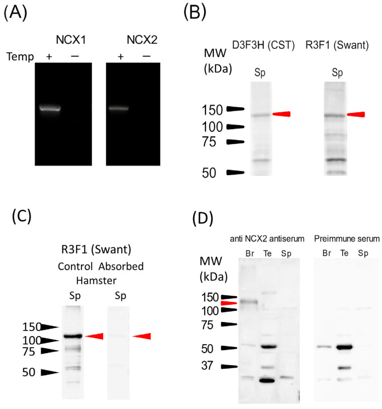
Previous studies demonstrated that hamster sperm hyperactivation is suppressed by extracellular Na+ by lowering intracellular Ca2+ levels, and Na+/Ca2+-exchanger (NCX) specific inhibitors canceled the suppressive effects of extracellular Na+. These results suggest the involvement of NCX in the regulation of hyperactivation. However, direct evidence of the presence and functionality of NCX in hamster spermatozoa is still lacking. This study aimed to reveal that NCX is present and is functional in hamster spermatozoa. First, NCX1 and NCX2 transcripts were detected via RNA-seq analyses of hamster testis mRNAs, but only the NCX1 protein was detected. Next, NCX activity was determined by measuring the Na+-dependent Ca2+ influx using the Ca2+ indicator Fura-2. The Na+-dependent Ca2+ influx was detected in hamster spermatozoa, notably in the tail region. The Na+-dependent Ca2+ influx was inhibited by the NCX inhibitor SEA0400 at NCX1-specific concentrations. NCX1 activity was reduced after 3 h of incubation in capacitating conditions. These results, together with authors' previous study, showed that hamster spermatozoa possesses functional NCX1 and that its activity was downregulated upon capacitation to trigger hyperactivation. This is the first study to successfully reveal the presence of NCX1 and its physiological function as a hyperactivation brake.

Keywords: NCX1; Na+/Ca2+ exchanger; sperm capacitation; sperm hyperactivation.


Conflict of interest statement

The authors declare no conflict of interest.

Figures

Figure 1
Figure 1
The expression of NCXs in hamster spermatozoa. The expression of NCX1 and NCX2 was examined via RT–PCR using hamster testis total RNA (A), and Western blotting using microsomal protein preparations from hamster spermatozoa (BD). (A) The expression of NCX1 and NCX2 mRNA in hamster testes was investigated using conventional RT–PCR. PCR products without templates were used as the negative control. (B) The expression of NCX1 protein in hamster spermatozoa was investigated via Western blotting using two different antibodies (D3F3H from CST and R3F1 from Swant) against NCX1. (C) The R3F1 antibody was preabsorbed by blocking peptide, and Western blotting was performed to validate the specificity of the antibody. (D) The expression of NCX2 protein in hamster spermatozoa and testes were investigated using custom rabbit anti–NCX2 antiserum. Hamster brain samples were used as a positive control. The results of antiserum (left) and preimmune serum (right) are shown side by side. The amount of protein loaded was 10 µg/well. Red arrowheads indicate a positive band. The molecular weights in kDa are shown on the left side.
Figure 2
Figure 2
The region-specific measurement of NCX activity in hamster. The schematics of how NCX activity measurement was carried out (AC) are shown, as well as the results of the measurements (D). The region of interest (ROI) was set as (A). (B): Na+-dependent Ca2+ influx was initiated via medium replacement (10–15 s), with the change in intracellular Ca2+ levels monitored by the ratio of Fura2. (C) The ratios of Fura2 in (B) 15 s after medium replacement were extracted and enlarged. The slope of each signal was defined as NCX activity. (D) The measured NCX activity (ratio/s) in head, midpiece, and principal piece are represented by a dot plot. Bars on the right represent mean ± S.D. A total of twenty-nine sperm cells from five individuals were assessed. Asterisks indicate significant differences among them (p < 0.05).
Figure 3
Figure 3
The effect of NCX inhibitor SEA0400 on Na+-dependent Ca2+ uptake by hamster spermatozoa. The effects of NCX inhibitor, SEA0400, on the Na+-dependent Ca2+ uptake were investigated. (AC) Both overall time-course changes (a) and 15–25 s after the medium replacement (b) of Fura-2 ratio in the presence of 0 (A), 1 (B), and 50 µM (C) of SEA0400 were shown. (DF) The measured NCX activity in head (D), midpiece (E), and principal piece (F) were normalized by those with an absence of SEA0400 and is represented by bar chart. Data are represented as mean ± S. D. Data were analyzed via a one-way ANOVA and the Tukey–Kramer post hoc test. Asterisks indicate significant differences among results (p < 0.05). A total of 26 cells was used to serve as the control, 30 cells for 1 µmol/L SEA0400, and 29 cells for 50 µmol/L SEA0400; all samples analyzed were taken from three individuals.
Figure 4
Figure 4
Capacitation-dependent change of NCX1 activity. (A) The change of NCX1 activity upon capacitation was investigated by measuring the NCX1 activity before (0 h) and 3 h after incubation (3 h) in capacitating conditions. (B,C) Basal intracellular Ca2+ levels of sperm before medium replacement were evaluated by Fura-2 ratio in the mTALP medium (B) or in the BSS medium (C) to confirm whether capacitation changed basal intracellular Ca2+ level during NCX activity measurement. (D) The NCX1 activity values (ratio/s) were calibrated by dividing them via the basal Fura-2 ratio in BSS medium (see (C)) and were normalized via the respective NCX1 activity values at 0 h in each region. Data are expressed as mean ± S.D. Data were analyzed using an (unpaired) Student’s t-test. Asterisks indicate significant differences among results (p < 0.05). A total of 29 cells for 0 h and 25 cells for 3 h from 5 individuals were analyzed in (A,C,D), and a total of 28 cells for 0 h and 22 cells for 3 h is analyzed in (B). (E) Protein levels of NCX1 before (0 h) and after (3 h) capacitation were investigated by Western blotting using microsomal preparations as a sample. The amount of protein loaded was 4 µg/well. Red arrowheads indicate a positive band. The molecular weights are shown on the left side. (F) The densitometry analysis of NCX1 bands was performed using Image J software for the comparison of NCX1 levels between 0 and 3 h. There was no significant difference between the results (p = 0.345). Data are expressed as mean ± S.D. n = 3.
Figure 5
Figure 5
PIP2 level was decreased during capacitation. Hamster spermatozoa were incubated in capacitating condition for 0 h or 3 h, then the cells were fixed and labeled with anti-PIP2 antibodies and Alexa Fluor 488-conjugated secondary antibodies. The labeled cells were analyzed through flow cytometry to determine sperm PIP2 levels. (A) Typical histograms were represented. The histogram with dotted line represents the negative control (secondary antibody only). A bar on the top left represents the population of PIP2-negative cells. (B) Percentages of PIP2-positive cells of four individuals, as analyzed in (A), and are represented by bar charts and dot plots. Data are expressed as mean ± S.D. Asterisks indicate the significant difference between results (p = 0.01).
Figure 6
Figure 6
Schematics of hamster sperm hyperactivation regulation by NCX1. In an uncapacitated spermatozoa, NCX1 is active and extrudes intracellular Ca2+, which it presumably entered through the CatSper channel, thereby suppressing hyperactivation and remaining in an activation state. In capacitated spermatozoa, NCX1 activity is downregulated by a decrease in PIP2 levels and inactive. Thus, Ca2+ ions entered through the CatSper channel are not extruded. This causes an increase in intracellular Ca2+ levels ([Ca2+]i), leading to an expression of hyperactivated motility.

References

    1. Yanagimachi R. Mammalian fertilization. In: Knobil E., Neill. J.D., editors. Physiology of Reproduction. 2nd ed. Raven Press; New York, NY, USA: 1994. pp. 189–317.
    1. Yanagimachi R. The movement of golden hamster spermatozoa before and after capacitation. J. Reprod. Fert. 1970;23:193–196. doi: 10.1530/jrf.0.0230193. - DOI - PubMed
    1. Suarez S.S., Katz D.F., Owen D.H., Andrew J.B., Powell R.L. Evidence for the function of hyperactivated motility in sperm. Biol. Reprod. 1991;44:375–381. doi: 10.1095/biolreprod44.2.375. - DOI - PubMed
    1. Suarez S.S., Dai X. Hyperactivation enhances mouse sperm capacity for penetrating viscoelastic media. Biol. Reprod. 1992;46:686–691. doi: 10.1095/biolreprod46.4.686. - DOI - PubMed
    1. Quill T.A., Sugden S., Rossi K., Doolittle L., Hammer R., Garbers D. Hyperactivated sperm motility driven by CatSper2 is required for fertilization. Proc. Natl. Acad. Sci. USA. 2003;100:14869–14874. doi: 10.1073/pnas.2136654100. - DOI - PMC - PubMed

LinkOut - more resources