Review
. 2023 May 19;24(10):9006.
doi: 10.3390/ijms24109006.

Extracellular-Vesicle-Based Therapeutics in Neuro-Ophthalmic Disorders

Affiliations
Review

Extracellular-Vesicle-Based Therapeutics in Neuro-Ophthalmic Disorders

Hamed Massoumi et al. Int J Mol Sci. .

Abstract

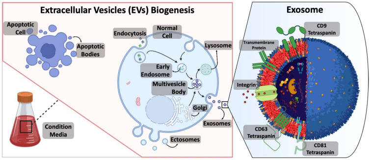
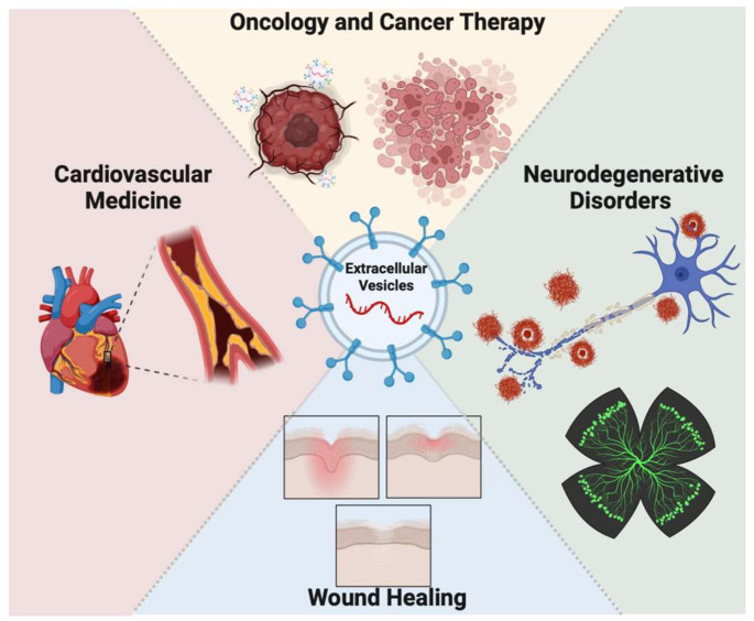
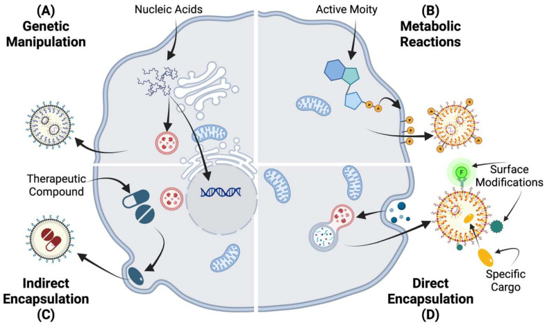
Extracellular vesicles (EVs) have been recognized as promising candidates for developing novel therapeutics for a wide range of pathologies, including ocular disorders, due to their ability to deliver a diverse array of bioactive molecules, including proteins, lipids, and nucleic acids, to recipient cells. Recent studies have shown that EVs derived from various cell types, including mesenchymal stromal cells (MSCs), retinal pigment epithelium cells, and endothelial cells, have therapeutic potential in ocular disorders, such as corneal injury and diabetic retinopathy. EVs exert their effects through various mechanisms, including promoting cell survival, reducing inflammation, and inducing tissue regeneration. Furthermore, EVs have shown promise in promoting nerve regeneration in ocular diseases. In particular, EVs derived from MSCs have been demonstrated to promote axonal regeneration and functional recovery in various animal models of optic nerve injury and glaucoma. EVs contain various neurotrophic factors and cytokines that can enhance neuronal survival and regeneration, promote angiogenesis, and modulate inflammation in the retina and optic nerve. Additionally, in experimental models, the application of EVs as a delivery platform for therapeutic molecules has revealed great promise in the treatment of ocular disorders. However, the clinical translation of EV-based therapies faces several challenges, and further preclinical and clinical studies are needed to fully explore the therapeutic potential of EVs in ocular disorders and to address the challenges for their successful clinical translation. In this review, we will provide an overview of different types of EVs and their cargo, as well as the techniques used for their isolation and characterization. We will then review the preclinical and clinical studies that have explored the role of EVs in the treatment of ocular disorders, highlighting their therapeutic potential and the challenges that need to be addressed for their clinical translation. Finally, we will discuss the future directions of EV-based therapeutics in ocular disorders. Overall, this review aims to provide a comprehensive overview of the current state of the art of EV-based therapeutics in ophthalmic disorders, with a focus on their potential for nerve regeneration in ocular diseases.

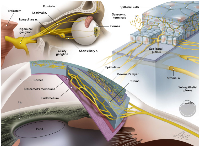
Keywords: cornea; exosomes; extracellular vesicles; nerve innervation; neuro-ophthalmic; ocular surfaces.


Conflict of interest statement

The authors declare no competing financial or nonfinancial conflict of interest.

Figures

Figure 1
Figure 1
Schematic representation of corneal layers and sensory nerve innervation in the human eye. The cellular layers of epithelium, stroma, and endothelium are distinguished from each other by two acellular membranes of Bowman’s and Descemet’s membranes. Sensory neuron innervation is schematically presented in the cornea inlet. The sensory nerves are originated in the trigeminal ganglion and penetrate to the cornea through the stroma layer (stromal nerves). As the nerve branches climb up to the surface of the cornea, they start dividing into thinner neurites (subepithelial plexus) right before they traverse through Bowman’s membrane and divide into even thinner neurites through basal epithelium layer and finally, end in sensory terminals in the epithelium, which is the outermost layer of the cornea.
Figure 2
Figure 2
Biogenesis of EVs from apoptotic bodies or healthy cells incubated in a condition media. It is possible to predominantly classify EVs into three groups. Apoptotic bodies that are cell blebs produced at the time of apoptosis. Ectosomes, also known as microvesicles, are produced by the plasma membrane’s outward budding and dissociation. Exosomes are intracellular products of the endocytic membrane transport pathway interrupted by the fusion of multivesicular bodies with the plasma membrane. As depicted in the magnified inlet, exosomes are phospholipid bilayer containers encapsulating a variety of components (i.e., proteins, lipids, nucleic acids, metabolites, etc.) with multiple membrane receptors, proteins, and biomarkers.
Figure 3
Figure 3
Therapeutic applications of EVs in clinical and preclinical studies published in the past decade are mostly focused on oncology and cancer therapy, cardiovascular medicine development, neurodegenerative diseases, and wound healing. The applications of EVs expand from diagnostic tools to novel therapeutics and can cover a wide range of organs and tissues.
Figure 4
Figure 4
Engineering approaches in the modification of EVs can be categorized into four categories: (A) Coding and noncoding oligonucleotides can be introduced into cells through genetic engineering. Their genetic changes can impact the expression of different nucleic acid expressions in the EVs’ cargo. (B) Metabolic labeling is used for introducing non-native moieties into cells. In this technique, metabolite replicas are integrated into cell biosynthesis. (C) Exogenous materials can be introduced to the cells and indirectly load the secreting EVs. (D) Direct EV modification is to permeabilize the vesicle membrane to enable the active loading of EVs. The vesicle membrane itself can potentially be the site of chemical reactions.

References

    1. Gehring W.J. The evolution of vision. Wiley Interdiscip Rev. Dev. Biol. 2014;3:1–40. doi: 10.1002/wdev.96. - DOI - PubMed
    1. Khosravimelal S., Mobaraki M., Eftekhari S., Ahearne M., Seifalian A.M., Gholipourmalekabadi M. Hydrogels as Emerging Materials for Cornea Wound Healing. Small. 2021;17:e2006335. doi: 10.1002/smll.202006335. - DOI - PubMed
    1. Yang A.Y., Chow J., Liu J. Corneal Innervation and Sensation: The Eye and Beyond. Yale J. Biol. Med. 2018;91:13–21. - PMC - PubMed
    1. Pham T.L., Bazan H.E. Docosanoid signaling modulates corneal nerve regeneration: Effect on tear secretion, wound healing, and neuropathic pain. J. Lipid Res. 2021;62:100033. doi: 10.1194/jlr.TR120000954. - DOI - PMC - PubMed
    1. Bouheraoua N., Fouquet S., Marcos-Almaraz M.T., Karagogeos D., Laroche L., Chédotal A. Genetic Analysis of the Organization, Development, and Plasticity of Corneal Innervation in Mice. J. Neurosci. 2019;39:1150–1168. doi: 10.1523/JNEUROSCI.1401-18.2018. - DOI - PMC - PubMed

LinkOut - more resources