. 2023 May 19;24(10):9028.
doi: 10.3390/ijms24109028.

Interaction of Bacteria, Immune Cells, and Surface Topography in Periprosthetic Joint Infections

Affiliations

Interaction of Bacteria, Immune Cells, and Surface Topography in Periprosthetic Joint Infections

Cristina Belgiovine et al. Int J Mol Sci. .

Abstract

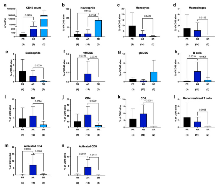
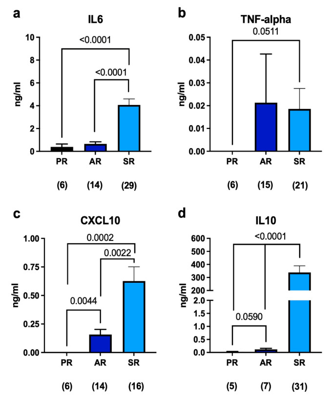
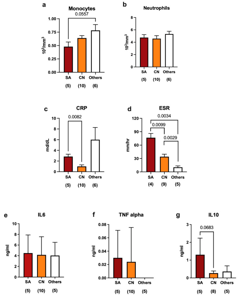
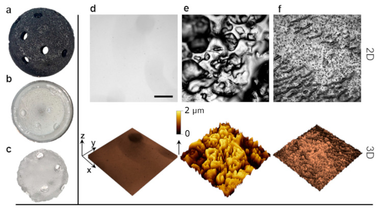
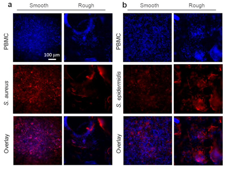
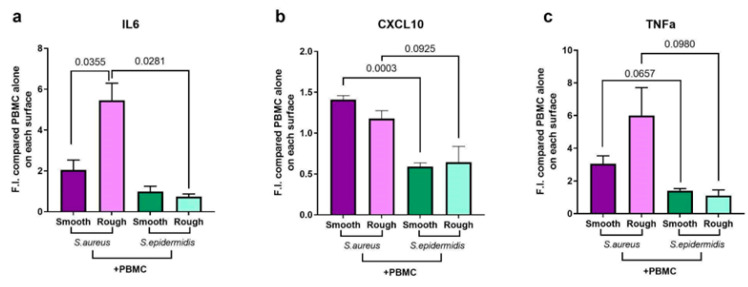
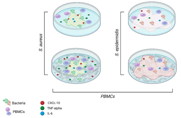
The incidence of periprosthetic joint infections (PJIs) is ~2% of total procedures and it is expected to rise due to an ageing population. Despite the large burden PJI has on both the individual and society, the immune response to the most commonly isolated pathogens, i.e., Staphylococcus aureus and Staphylococcus epidermidis, remains incompletely understood. In this work, we integrate the analysis of synovial fluids from patients undergoing hip and knee replacement surgery with in-vitro experimental data obtained using a newly developed platform, mimicking the environment of periprosthetic implants. We found that the presence of an implant, even in patients undergoing aseptic revisions, is sufficient to induce an immune response, which is significantly different between septic and aseptic revisions. This difference is confirmed by the presence of pro- and anti-inflammatory cytokines in synovial fluids. Moreover, we discovered that the immune response is also dependent on the type of bacteria and the topography of the implant surface. While S. epidermidis seems to be able to hide better from the attack of the immune system when cultured on rough surfaces (indicative of uncemented prostheses), S. aureus reacts differently depending on the contact surface it is exposed to. The experiments we performed in-vitro also showed a higher biofilm formation on rough surfaces compared to flat ones for both species, suggesting that the topography of the implant could influence both biofilm formation and the consequent immune response.

Keywords: immune response; periprosthetic joint infection (PJI); staphylococcal biofilms; surface topography.


Conflict of interest statement

The authors declare no conflict of interest.

Figures

Figure 1
Figure 1
FACS analysis of periprosthetic leukocytes derived from patients undergoing either primary replacement (PR), aseptic revision (AR), or septic revision (SR). (a) CD45 cell count was performed using beads derived from Leukocounts kit (Becton and Dickinson, Franklin Lakes, NJ, USA) on a subset of samples. (bl) Live cells were selected and the percentage was calculated on CD45-positive viable cells. (m,n) Analysis of T-cell activation was performed on different patients using CD69 as marker. The number below each group represents the number of samples analyzed. Histograms represent the mean ± standard deviation. Statistical analysis: unpaired t-test with Welch’s correction.
Figure 2
Figure 2
(ad) ELISA quantification of soluble mediators in the synovial fluid of patients undergoing either primary replacement or revision surgeries. The number below each group represents the number of samples analyzed. Histograms represent the mean ± standard deviation. Statistical analysis: unpaired t-test with Welch’s correction.
Figure 3
Figure 3
Immune modulation in response to different bacterial species in septic revisions. (ad) Blood-circulating monocytes, neutrophils, C-reactive protein (CRP), and erythrocyte sedimentation rate (ESR) indicate the status of infection at systemic levels. (eg) ELISA quantification of IL-6, TNF alpha, and IL-10 in the synovial fluids of patients with joints contaminated by different bacterial species. The number below each group represents the number of samples analyzed. Histograms represent the mean ± standard deviation. Statistical analysis: unpaired t-test with Welch’s correction.
Figure 4
Figure 4
Fabrication of the PJI in vitro platform. (a) Commercial trabecular metal (TM) tantalum acetabular cup. (b) Negative replica of the cup in hard polyurethane binary resin. (c) Positive polydimethilsiloxane (PDMS) replica employed as a model substrate. (df) Surface characterization of different surface topographies. Phase contrast (PH) optical microscopy images and 3D topography projection of PDMS-replicated smooth and rough surfaces (d,e) compared with a cemented reference sample (f). Scale bar is 500 µm.
Figure 5
Figure 5
Fluorescence optical microscopy analysis for the in vitro model topographies in the presence of (a) PBMCs and S. aureus and (b) PBMCs and S. epidermidis. PBMCs were stained by means of CFSE CellTrace emitting at 517 nm (blue channel), whereas for Staph. spp., a CMPTX Cell Tracker emitting at 602 nm (red channel) was used. Panels (a,b) report single blue and red channels and an overlay of the two merged channels.
Figure 6
Figure 6
Biofilm quantification by crystal violet staining. (a,b) Biofilm quantification assay at 24 h (a) and 48 h (b) for S. aureus and S. epidermidis on smooth and rough surfaces. Both bacteria preferentially colonize rough surfaces, although, at 48 h, S. epidermidis biofilm production appears higher in general compared to that of S. aureus on both surfaces. (c,d) Biofilm quantification assay at 24 h and 48 h for S. aureus or S. epidermidis in the presence of PBMCs. At 24 h (c), bacterial growth is prominent and not influenced by immune cells. At 48 h (d), on rough surfaces, a reduction in biofilm production is observed indicating a response from immune cells to microbial proliferation. Statistical analysis: unpaired t-test with Welch’s correction.
Figure 7
Figure 7
ELISA quantification in the co-culture of PBMCs and bacteria compared to the ELISA quantification of PBMCs cultured alone on different textured surfaces. We tested the release of (a) IL-6, (b) CXCL10, and (c) TNF-alpha in the supernatant. The columns represent the mean ± standard deviation of three independent donors. Statistical analysis: unpaired t-test with Welch’s correction.
Figure 8
Figure 8
Schematic illustration summarizing the key features from the in vitro experimental findings. Created with BioRender.com.

References

    1. Nodzo S.R., Boyle K.K., Spiro S., Nocon A.A., Miller A.O., Westrich G.H. Success Rates, Characteristics, and Costs of Articulating Antibiotic Spacers for Total Knee Periprosthetic Joint Infection. Knee. 2017;24:1175–1181. doi: 10.1016/j.knee.2017.05.016. - DOI - PubMed
    1. Patel R. Periprosthetic Joint Infection. N. Engl. J. Med. 2023;388:251–262. doi: 10.1056/NEJMra2203477. - DOI - PubMed
    1. Zimmerli W., Trampuz A., Ochsner P.E. Prosthetic-Joint Infections. N. Engl. J. Med. 2004;351:1645–1654. doi: 10.1056/NEJMra040181. - DOI - PubMed
    1. Oliveira W.F., Silva P.M.S., Silva R.C.S., Silva G.M.M., Machado G., Coelho L.C.B.B., Correia M.T.S. Staphylococcus Aureus and Staphylococcus Epidermidis Infections on Implants. J. Hosp. Infect. 2018;98:111–117. doi: 10.1016/j.jhin.2017.11.008. - DOI - PubMed
    1. Arciola C.R., Campoccia D., Montanaro L. Implant Infections: Adhesion, Biofilm Formation and Immune Evasion. Nat. Rev. Microbiol. 2018;16:397–409. doi: 10.1038/s41579-018-0019-y. - DOI - PubMed

MeSH terms