

Cardiac Magnetic Resonance in HCM Phenocopies: From Diagnosis to Risk Stratification and Therapeutic Management
- PMID: 37240587
- PMCID: PMC10218866
- DOI: 10.3390/jcm12103481
Cardiac Magnetic Resonance in HCM Phenocopies: From Diagnosis to Risk Stratification and Therapeutic Management
Abstract
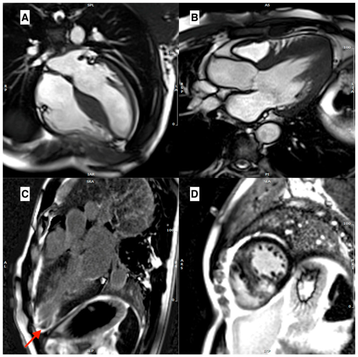
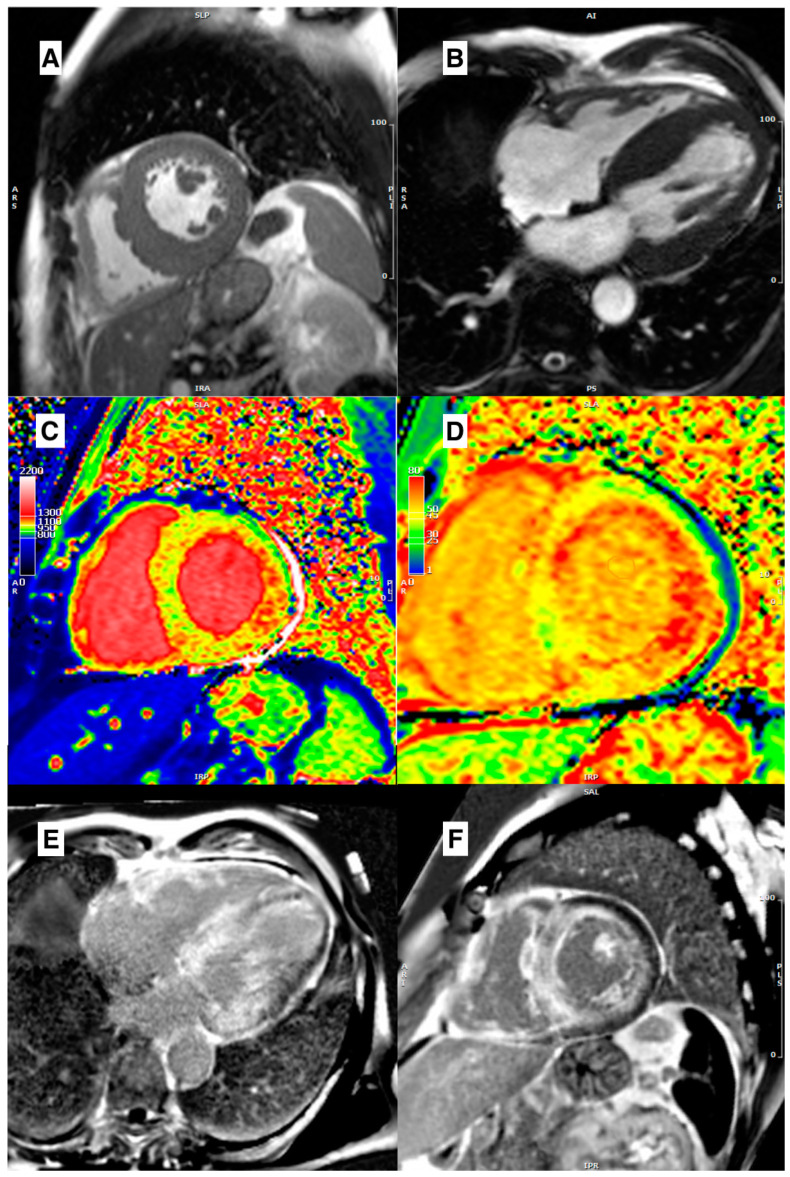
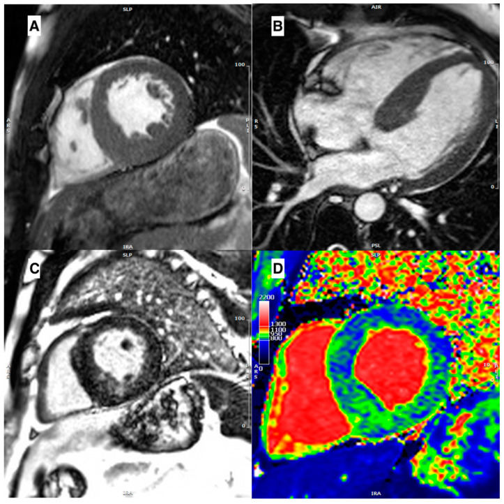
Hypertrophic cardiomyopathy (HCM) is a genetic heart disease characterized by the thickening of the heart muscle, which can lead to symptoms such as chest pain, shortness of breath, and an increased risk of sudden cardiac death. However, not all patients with HCM have the same underlying genetic mutations, and some have conditions that resemble HCM but have different genetic or pathophysiological mechanisms, referred to as phenocopies. Cardiac magnetic resonance (CMR) imaging has emerged as a powerful tool for the non-invasive assessment of HCM and its phenocopies. CMR can accurately quantify the extent and distribution of hypertrophy, assess the presence and severity of myocardial fibrosis, and detect associated abnormalities. In the context of phenocopies, CMR can aid in the differentiation between HCM and other diseases that present with HCM-like features, such as cardiac amyloidosis (CA), Anderson-Fabry disease (AFD), and mitochondrial cardiomyopathies. CMR can provide important diagnostic and prognostic information that can guide clinical decision-making and management strategies. This review aims to describe the available evidence of the role of CMR in the assessment of hypertrophic phenotype and its diagnostic and prognostic implications.
Keywords: cardiac magnetic resonance; hypertrophic cardiomyopathy; phenocopies.
Conflict of interest statement
The authors declare no conflict of interest.
Figures
References
-
- Elliott P., Andersson B., Arbustini E., Bilinska Z., Cecchi F., Charron P., Dubourg O., Kühl U., Maisch B., McKenna W.J., et al. Classification of the cardiomyopathies: A position statement from the european society of cardiology working group on myocardial and pericardial diseases. Eur. Heart J. 2007;29:270–276. doi: 10.1093/eurheartj/ehm342. - DOI - PubMed
-
- Licordari R., Minutoli F., Cappelli F., Micari A., Colarusso L., Di Paola F.A.F., Campisi M., Recupero A., Mazzeo A., Di Bella G. Mid-basal left ventricular longitudinal dysfunction as a prognostic marker in mutated transthyretin-related cardiac amyloidosis. Vessel. Plus. 2022;6:12. doi: 10.20517/2574-1209.2021.86. - DOI
Publication types
LinkOut - more resources
Full Text Sources
