. 2023 May 17;12(10):3514.
doi: 10.3390/jcm12103514.

The Prognostic and Diagnostic Value of [18F]FDG PET/CT in Untreated Laryngeal Carcinoma

Affiliations

The Prognostic and Diagnostic Value of [18F]FDG PET/CT in Untreated Laryngeal Carcinoma

Akram Al-Ibraheem et al. J Clin Med. .

Abstract

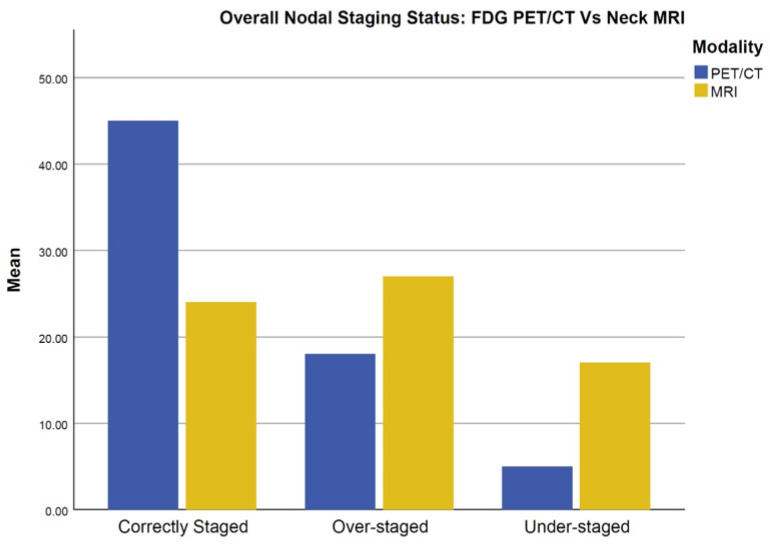
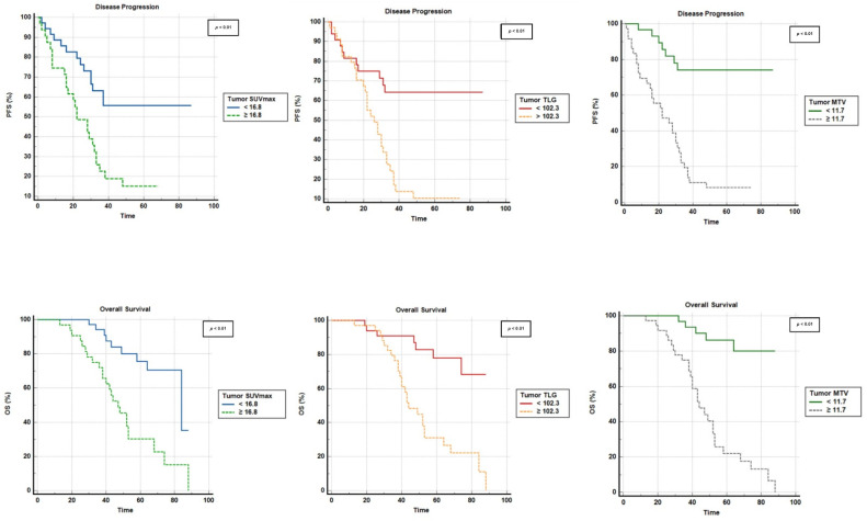
This study aims to determine the diagnostic accuracy of staging PET/CT and neck MRI in patients with laryngeal carcinoma and to assess the value of PET/CT in predicting progression-free survival (PFS) and overall survival (OS). Sixty-eight patients who had both modalities performed before treatment between 2014 and 2021 were included in this study. The sensitivity and specificity of PET/CT and MRI were evaluated. PET/CT had 93.8% sensitivity, 58.3% specificity, and 75% accuracy for nodal metastasis, whereas MRI had 68.8%, 61.1%, and 64.7% accuracy, respectively. At a median follow-up of 51 months, 23 patients had developed disease progression and 17 patients had died. Univariate-survival analysis revealed all utilized PET parameters as significant prognostic factors for OS and PFS (p-value < 0.03 each). In multivariate analysis, metabolic-tumor volume (MTV) and total lesion glycolysis (TLG) predicted better PFS (p-value < 0.05 each). In conclusion, PET/CT improves the accuracy of nodal staging in laryngeal carcinoma over neck MRI and adds to the prognostication of survival outcomes through the use of several PET metrics.

Keywords: MTV; PET/CT; TLG; [18F]FDG PET/CT vs. neck MRI; laryngeal squamous-cell carcinoma; metastatic lymph node; semiquantitative PET parameters.


Conflict of interest statement

The authors declare no conflict of interest.

Figures

Figure 1
Figure 1
(a) MIP; (b) axial PET/CT, and axial PET; (c) sagittal PET/CT images showing an intensely hypermetabolic malignant glottic-mass lesion (arrowheads) with direct invasion to left para-glottic space and thyroid cartilage. This was associated with a single hypermetabolic left level III cervical lymph node (arrows). The overall staging result was T4aN1M0, which is consistent with the post-operative biopsy.
Figure 2
Figure 2
A 49-year-old male patient underwent baseline neck MRI followed by [18F]FDG PET/CT for staging purposes. (a) Sagittal PET/CT and sagittal T1 MRI images showed a large lobulated transglottic-mass lesion with an extra-laryngeal extension that was highly [18F]FDG avid and heterogeneous on MRI (arrowheads). (b) Axial PET/CT and axial T2 MR images showed a prominent left level IIa cervical lymph node that was hypermetabolic and enhanced by MRI (arrows), suggesting metastases. Other right level IIa and left III–IV lymph nodes were heterogeneously enhanced but non-[18F]FDG avid (curved arrows). [18F]FDG PET/CT nodal staging outperformed MRI and matched the biopsy results (N1 nodal disease).
Figure 3
Figure 3
Overall nodal-staging status: [18F]FDG PET/CT vs. neck MRI.
Figure 4
Figure 4
Kaplan–Meir survival curves according to baseline tumor SUVmax, TLG, and MTV.

References

    1. Steuer C.E., El-Deiry M., Parks J.R., Higgins K.A., Saba N.F. An update on larynx cancer. CA Cancer J. Clin. 2017;67:31–50. doi: 10.3322/caac.21386. - DOI - PubMed
    1. Almadori G., Bussu F., Cadoni G., Galli J., Paludetti G., Maurizi M. Molecular markers in laryngeal squamous cell carcinoma: Towards an integrated clinicobiological approach. Eur. J. Cancer. 2005;41:683–693. doi: 10.1016/j.ejca.2004.10.031. - DOI - PubMed
    1. Forastiere A.A., Ismaila N., Lewin J.S., Nathan C.A., Adelstein D.J., Eisbruch A., Fass G., Fisher S.G., Laurie S.A., Le Q.T., et al. Use of Larynx-Preservation Strategies in the Treatment of Laryngeal Cancer: American Society of Clinical Oncology Clinical Practice Guideline Update. J. Clin. Oncol. 2018;36:1143–1169. doi: 10.1200/JCO.2017.75.7385. - DOI - PubMed
    1. Kyzas P.A., Evangelou E., Denaxa-Kyza D., Ioannidis J.P. 18F-fluorodeoxyglucose positron emission tomography to evaluate cervical node metastases in patients with head and neck squamous cell carcinoma: A meta-analysis. J. Natl. Cancer Inst. 2008;100:712–720. doi: 10.1093/jnci/djn125. - DOI - PubMed
    1. Yongkui L., Jian L., Wanghan, Jingui L. 18FDG-PET/CT for the detection of regional nodal metastasis in patients with primary head and neck cancer before treatment: A meta-analysis. Surg. Oncol. 2013;22:e11–e16. doi: 10.1016/j.suronc.2013.02.002. - DOI - PubMed

LinkOut - more resources