

The Evaluation of Cartilage Regeneration Efficacy of Three-Dimensionally Biofabricated Human-Derived Biomaterials on Knee Osteoarthritis: A Single-Arm, Open Label Study in Egypt
- PMID: 37240918
- PMCID: PMC10222898
- DOI: 10.3390/jpm13050748
The Evaluation of Cartilage Regeneration Efficacy of Three-Dimensionally Biofabricated Human-Derived Biomaterials on Knee Osteoarthritis: A Single-Arm, Open Label Study in Egypt
Abstract
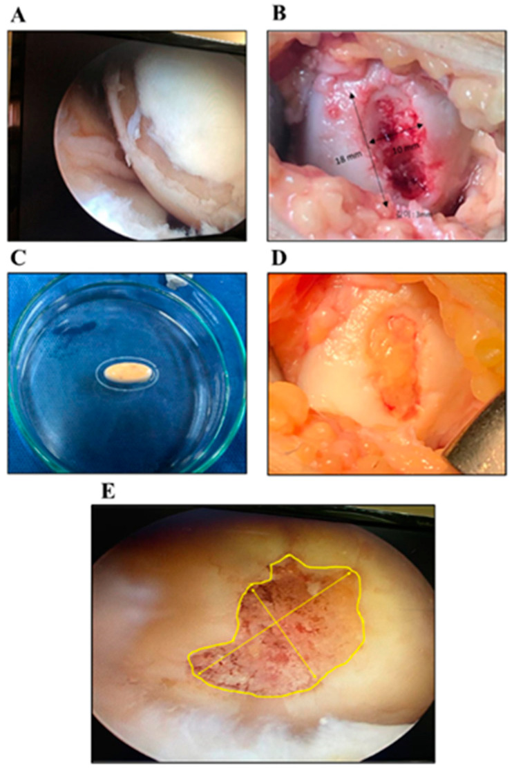
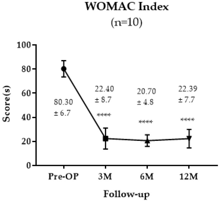
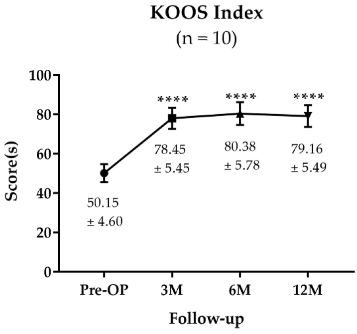
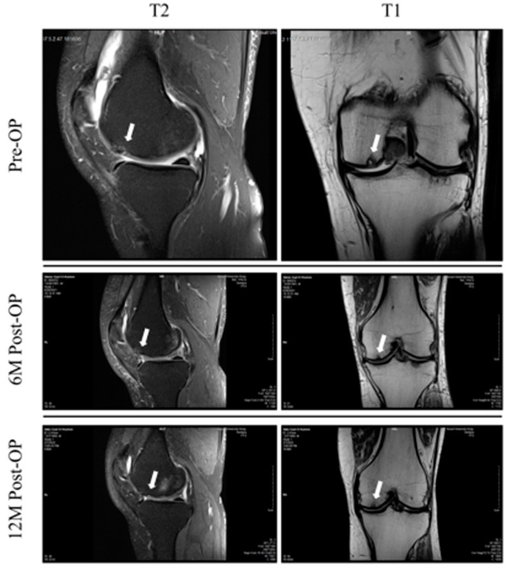
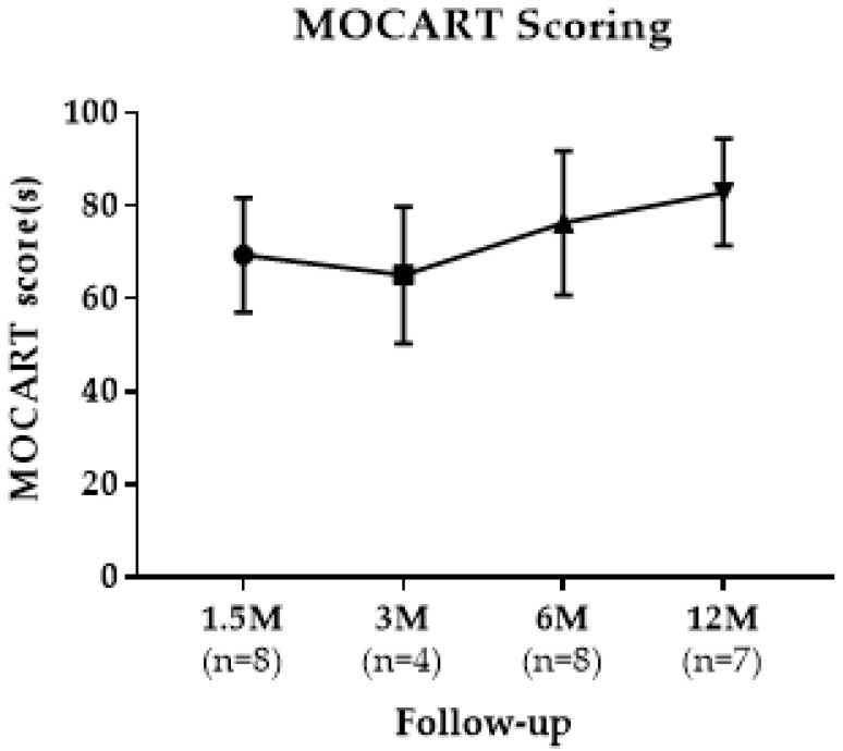
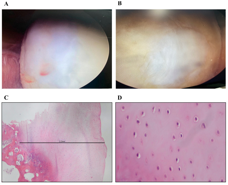
Full thickness cartilage defects in cases of knee osteoarthritis are challenging in nature and are difficult to treat. The implantation of three-dimensional (3D) biofabricated grafts into the defect site can be a promising biological one-stage solution for such lesions that can avoid different disadvantages of the alternative surgical treatment options. In this study, the short-term clinical outcome of a novel surgical technique that uses a 3D bioprinted micronized adipose tissue (MAT) graft for knee cartilage defects is assessed and the degree of incorporation of such graft types is evaluated via arthroscopic and radiological analyses. Ten patients received 3D bioprinted grafts consisting of MAT with an allogenic hyaline cartilage matrix on a mold of polycaprolactone, with or without adjunct high tibial osteotomy, and they were monitored until 12 months postoperatively. Clinical outcomes were examined with patient-reported scoring instruments that consisted of the Western Ontario and McMaster Universities Arthritis Index (WOMAC) score and the Knee Injury and Osteoarthritis Outcome Score (KOOS). The graft incorporation was assessed using the Magnetic Resonance Observation of Cartilage Repair Tissue (MOCART) score. At 12 months follow-up, cartilage tissue biopsy samples were taken from patients and underwent histopathological examination. In the results, at final follow-up, the WOMAC and KOOS scores were 22.39 ± 7.7 and 79.16 ± 5.49, respectively. All scores were significantly increased at final follow-up (p < 0.0001). MOCART scores were also improved to a mean of 82.85 ± 11.49, 12 months after operation, and we observed a complete incorporation of the grafts with the surrounding cartilage. Together, this study suggests a novel regeneration technique for the treatment of knee osteoarthritis patients, with less rejection response and better efficacy.
Keywords: 3D bioprinting; adipose tissue; allogenic hyaline cartilage; articular cartilage; knee; osteoarthritis.
Conflict of interest statement
The authors declare no conflict of interest.
Figures
References
-
- Gu X., Li C., Yin F., Yang G. Adipose-derived stem cells in articular cartilage regeneration: Current concepts and optimization strategies. Histol. Histopathol. 2018;33:639–653. - PubMed
LinkOut - more resources
Full Text Sources
