

Current Concepts in the Management of Primary Lymphedema
- PMID: 37241126
- PMCID: PMC10222882
- DOI: 10.3390/medicina59050894
Current Concepts in the Management of Primary Lymphedema
Abstract
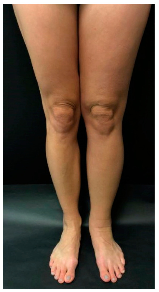
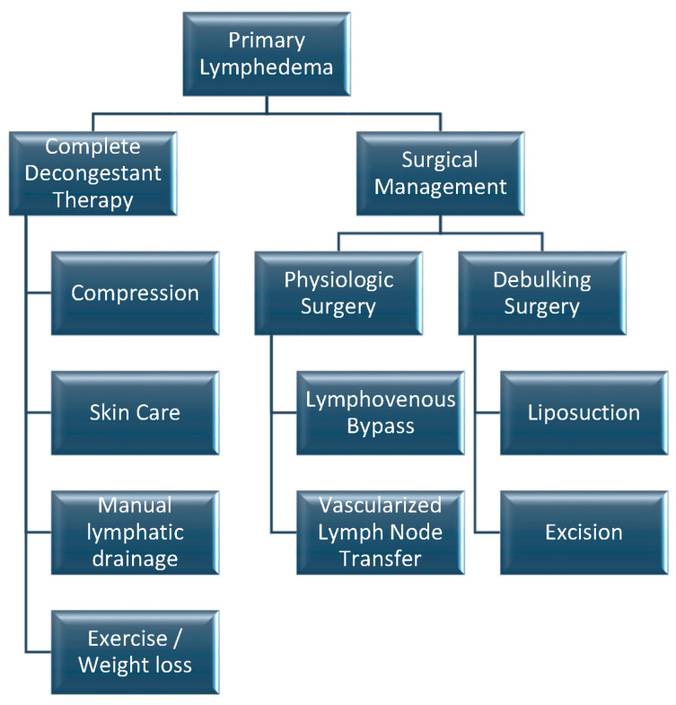
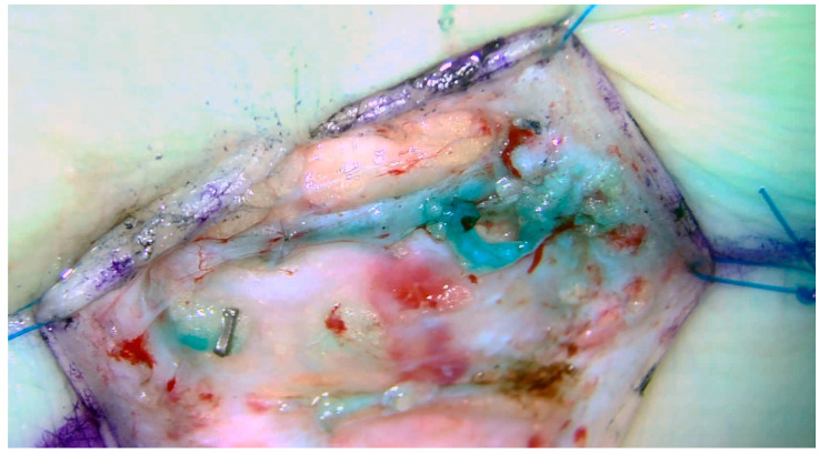
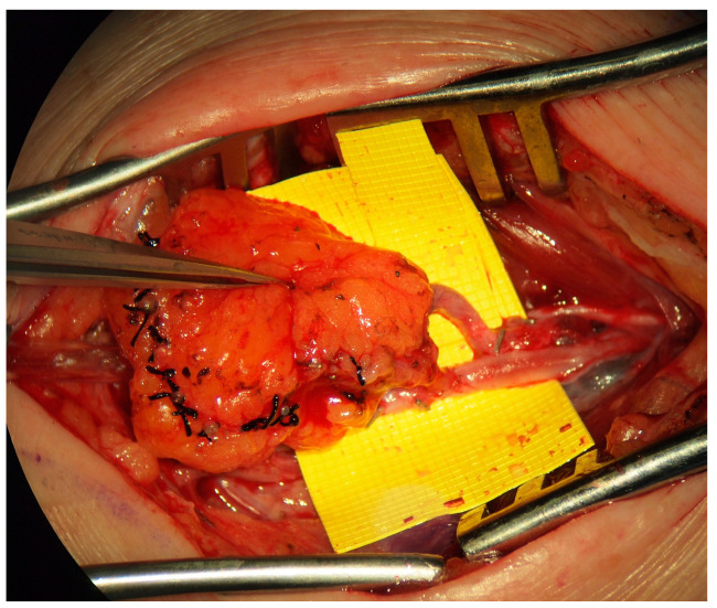
Primary lymphedema is a heterogeneous group of conditions encompassing all lymphatic anomalies that result in lymphatic swelling. Primary lymphedema can be difficult to diagnose, and diagnosis is often delayed. As opposed to secondary lymphedema, primary lymphedema has an unpredictable disease course, often progressing more slowly. Primary lymphedema can be associated with various genetic syndromes or can be idiopathic. Diagnosis is often clinical, although imaging can be a helpful adjunct. The literature on treating primary lymphedema is limited, and treatment algorithms are largely based on practice patterns for secondary lymphedema. The mainstay of treatment focuses on complete decongestive therapy, including manual lymphatic drainage and compression therapy. For those who fail conservative treatment, surgical treatment can be an option. Microsurgical techniques have shown promise in primary lymphedema, with both lymphovenous bypass and vascularized lymph node transfers demonstrating improved clinical outcomes in a few studies.
Keywords: congenital lymphedema; lymphovenous anastomosis; lymphovenous bypass; primary lymphedema; vascularized lymph node transfer.
Conflict of interest statement
The authors declare no conflict of interest.
Figures

References
-
- Vignes S., Albuisson J., Champion L., Constans J., Tauveron V., Malloizel J., Quere I., Simon L., Arrault M., Trevidic P., et al. Primary lymphedema French National Diagnosis and Care Protocol (PNDS; Protocole National de Diagnostic et de Soins) Orphanet J. Rare Dis. 2021;16:18. doi: 10.1186/s13023-020-01652-w. - DOI - PMC - PubMed
-
- Bellini C., Hennekam R.C. Clinical disorders of primary malfunctioning of the lymphatic system. Adv. Anat. Embryol. Cell Biol. 2014;214:187–204. - PubMed
Publication types
MeSH terms
LinkOut - more resources
Full Text Sources
Medical
