. 2023 May 15;28(10):4092.
doi: 10.3390/molecules28104092.

Biocytin-Labeling in Whole-Cell Recording: Electrophysiological and Morphological Properties of Pyramidal Neurons in CYLD-Deficient Mice

Affiliations

Biocytin-Labeling in Whole-Cell Recording: Electrophysiological and Morphological Properties of Pyramidal Neurons in CYLD-Deficient Mice

Shuyi Tan et al. Molecules. .

Abstract

Biocytin, a chemical compound that is an amide formed from the vitamin biotin and the amino acid L-lysine, has been used as a histological dye to stain nerve cells. Electrophysiological activity and morphology are two key characteristics of neurons, but revealing both the electrophysiological and morphological properties of the same neuron is challenging. This article introduces a detailed and easy-to-operate procedure for single-cell labeling in combination with whole-cell patch-clamp recording. Using a recording electrode filled with a biocytin-containing internal solution, we demonstrate the electrophysiological and morphological characteristics of pyramidal (PNs), medial spiny (MSNs) and parvalbumin neurons (PVs) in brain slices, where the electrophysiological and morphological properties of the same individual cell are elucidated. We first introduce a protocol for whole-cell patch-clamp recording in various neurons, coupled with the intracellular diffusion of biocytin delivered by the glass capillary of the recording electrode, followed by a post hoc procedure to reveal the architecture and morphology of biocytin-labeled neurons. An analysis of action potentials (APs) and neuronal morphology, including the dendritic length, number of intersections, and spine density of biocytin-labeled neurons, were performed using ClampFit and Fiji Image (ImageJ), respectively. Next, to take advantage of the techniques introduced above, we uncovered defects in the APs and the dendritic spines of PNs in the primary motor cortex (M1) of deubiquitinase cylindromatosis (CYLD) knock-out (Cyld-/-) mice. In summary, this article provides a detailed methodology for revealing the morphology as well as the electrophysiological activity of a single neuron that will have many applications in neurobiology.

Keywords: CYLD; action potential; biocytin; dendritic spine; single-cell labeling; whole-cell patch-clamp recording.


Conflict of interest statement

The authors declare no conflict of interest.

Figures

Figure 1
Figure 1
Illustration of experimental procedure. (A) Flow diagram of experimental procedures. (B) Schematic of AAV-hSyn-DIO-EGFP injection into the DLS of PV-Cre mice. (C) Schematic illustration of the areas of acute brain slices and neuron types targeted for whole-cell recording and biocytin infusion. (D) Neurons were initially identified by the shape of their somata using IR-DIC microscopy and were then recorded by whole-cell patch-clamping with a standard stimulation paradigm for the characterization of intrinsic electrical properties. (E) Slices were incubated with Streptavidin Alexa 594 (red) or Streptavidin Alexa 488 (green) for 24 h to reveal biocytin-loaded neurons under a confocal microscope. Maximum-intensity projections are shown of a biocytin-labeled PN, MSN and PV interneuron.
Figure 2
Figure 2
Representative traces and current-voltage curve of whole-cell recorded APs of PV interneurons and biocytin labeling of the same cells. (A,B) Representative single and current injection-induced APs of a PV interneuron. (C) Current-voltage (I-V) curve of PV interneurons (n = 9 neurons). (D) EGFP-positive PV neuron labeled with biocytin (red) to reveal its morphology (white arrow).
Figure 3
Figure 3
Representative traces and I-V curve of whole-cell recorded APs of MSNs and biocytin labeling of the same cells. (A,B) Representative single and current injection-induced APs of an MSN. (C) I-V curve of MSNs (n = 10 neurons). (D) Morphology of an MSN revealed by biocytin.
Figure 4
Figure 4
Representative traces and I-V curve of whole-cell recorded APs of PNs and biocytin labeling of the same cells. (A,B) Representative single and current injection-induced APs of a PN. (C) I-V curve of PNs (n = 12 neurons). (D) PN morphology revealed by biocytin.
Figure 5
Figure 5
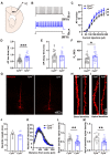
Apical and basal dendritic spine loss in PNs of Cyld−/− mice. (A) Diagram of the electrophysiological recording strategy in acute motor cortical slices. (B) Representative traces of AP firing of PNs of Cyld+/+ mice (black) and Cyld−/− mice (blue) in response to 150 pA (1000 ms duration) current injections. (C) Depolarizing current steps (+30 to +270 pA, 30 steps, 1000 ms duration) lead to an increase in AP frequency in Cyld−/− mice (n = 27 neurons from 11 Cyld+/+ mice, n = 22 neurons from 7 Cyld−/− mice). (DF) Bar graphs showing AP rise (D), decay time (E) and Rin (F) of PNs from Cyld+/+ and Cyld−/− mice (n = 26 neurons from 11 Cyld+/+ mice, n = 20 neurons from 7 Cyld−/− mice). (G) Representative image of an individual PN from Cyld+/+ or Cyld−/− mice loaded with biocytin via a patch microelectrode. PNs were imaged with a confocal microscope system, and images were used to analyze dendrite morphology and spine density. (H,I) Representative confocal images of basal dendrites (H) and apical dendrites (I) from Cyld+/+ and Cyld−/− mice. (J) Bar graphs showing dendritic length of PNs from Cyld+/+ and Cyld−/− mice. (K) Sholl analysis demonstrating that there is no difference between the genotypes in the neuronal dendritic arborization complexity of PNs. Points represent individual neurons (n = 7 neurons from three Cyld+/+ mice, n = 9 neurons from three Cyld−/− mice) in (J,K). (L,M) Bar graphs showing that basal (L) and apical (M) dendritic spine density of PNs decrease in Cyld−/− mice. Points represent single basal dendrites (n = 14 dendrites from Cyld+/+, n = 33 dendrites from Cyld−/−) in (L) and single apical dendrites (n = 11 dendrites from Cyld+/+, n = 23 dendrites from Cyld−/−) in (M). Data are presented as the mean ± SEM; * p < 0.05, ** p < 0.01, *** p < 0.001.
Figure 6
Figure 6
Decreased AHP duration and increased AHP decay in PNs of Cyld−/− mice. (A,B) Representative traces of AP firing of PNs in Cyld+/+ (A) and Cyld−/− mice (B) in response to 150 pA (1000 ms duration) current injection. Insets show expanded view of the spikes. (CE) Bar graphs of AHP duration (C), amplitude (D) and decay constant (E) (n = 29 neurons from 10 Cyld+/+ mice, n = 20 neurons from 5 Cyld−/− mice). Data are presented as the mean ± SEM; * p < 0.05, ** p < 0.01, *** p < 0.001.

References

    1. Hodapp A., Kaiser M.E., Thome C., Ding L., Rozov A., Klumpp M., Stevens N., Stingl M., Sackmann T., Lehmann N., et al. Dendritic axon origin enables information gating by perisomatic inhibition in pyramidal neurons. Science. 2022;377:1448–1452. doi: 10.1126/science.abj1861. - DOI - PubMed
    1. Kasai H., Ziv N.E., Okazaki H., Yagishita S., Toyoizumi T. Spine dynamics in the brain, mental disorders and artificial neural networks. Nat. Rev. Neurosci. 2021;22:407–422. doi: 10.1038/s41583-021-00467-3. - DOI - PubMed
    1. Peca J., Feliciano C., Ting J.T., Wang W., Wells M.F., Venkatraman T.N., Lascola C.D., Fu Z., Feng G. Shank3 mutant mice display autistic-like behaviours and striatal dysfunction. Nature. 2011;472:437–442. doi: 10.1038/nature09965. - DOI - PMC - PubMed
    1. Booker S.A., Domanski A.P.F., Dando O.R., Jackson A.D., Isaac J.T.R., Hardingham G.E., Wyllie D.J.A., Kind P.C. Altered dendritic spine function and integration in a mouse model of fragile X syndrome. Nat. Commun. 2019;10:4813. doi: 10.1038/s41467-019-11891-6. - DOI - PMC - PubMed
    1. Gouwens N.W., Sorensen S.A., Berg J., Lee C., Jarsky T., Ting J., Sunkin S.M., Feng D., Anastassiou C.A., Barkan E., et al. Classification of electrophysiological and morphological neuron types in the mouse visual cortex. Nat. Neurosci. 2019;22:1182–1195. doi: 10.1038/s41593-019-0417-0. - DOI - PMC - PubMed