. 2023 May 19;28(10):4190.
doi: 10.3390/molecules28104190.

Xanthophyll-Rich Extract of Phaeodactylum tricornutum Bohlin as New Photoprotective Cosmeceutical Agent: Safety and Efficacy Assessment on In Vitro Reconstructed Human Epidermis Model

Affiliations

Xanthophyll-Rich Extract of Phaeodactylum tricornutum Bohlin as New Photoprotective Cosmeceutical Agent: Safety and Efficacy Assessment on In Vitro Reconstructed Human Epidermis Model

Antonella Smeriglio et al. Molecules. .

Abstract

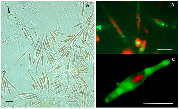
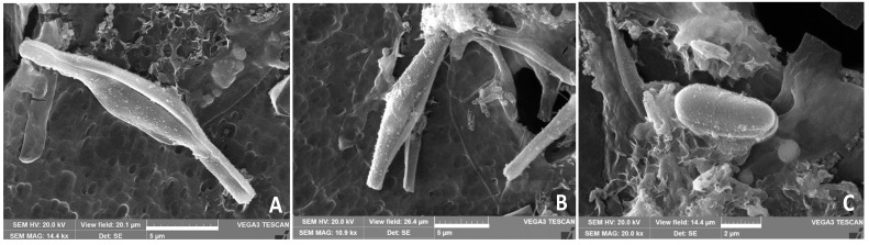
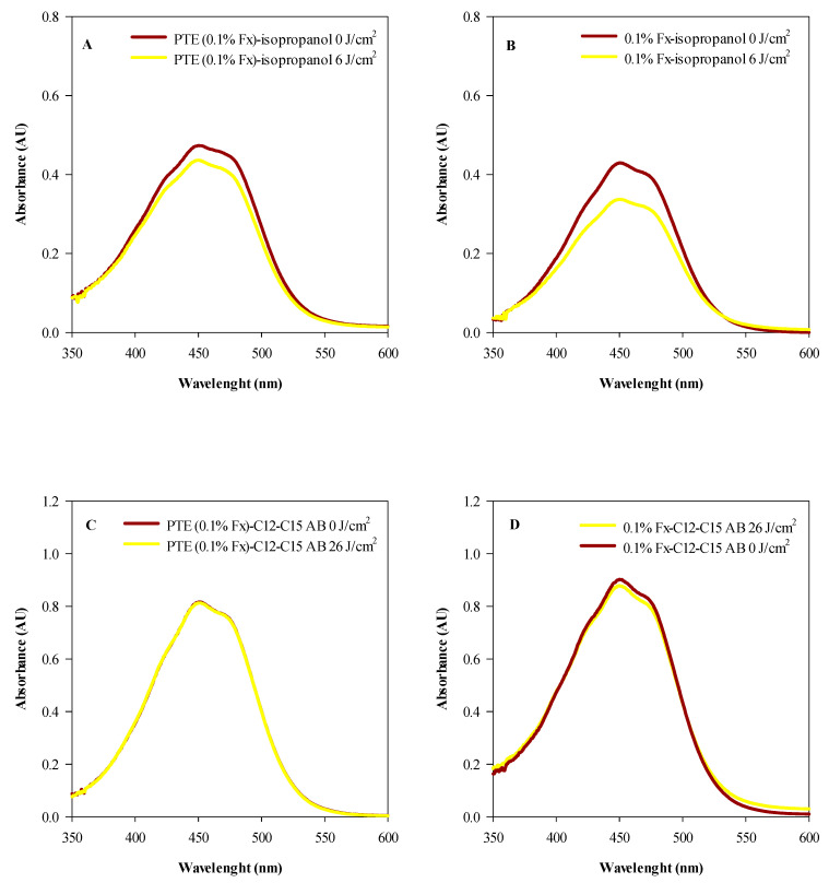
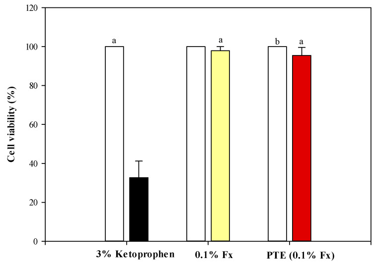
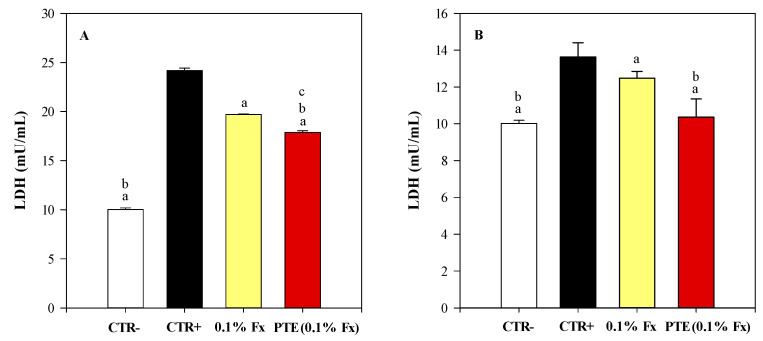
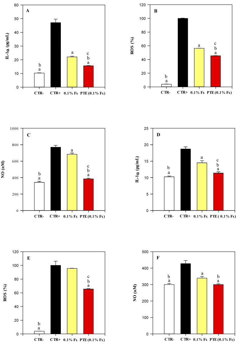
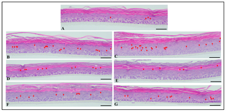
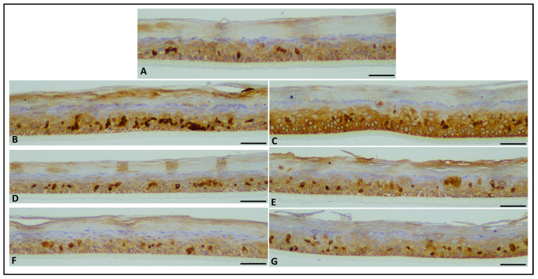
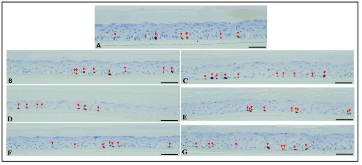
The nutritional and health properties of algae make them perfect functional ingredients for nutraceutical and cosmeceutical applications. In this study, the Phaeodactylum tricornutum Bohlin (Phaeodactylaceae), a pleiomorphic diatom commonly found in marine ecosystems, was investigated. The in vitro culture conditions used favoured the fusiform morphotype, characterized by a high accumulation of neutral lipids, as detected by fluorescence microscopy after BODIPY staining. These data were confirmed by HPLC-DAD-APCI-MS/MS analyses carried out on the ethanolic extract (PTE), which showed a high content of xanthophylls (98.99%), and in particular of fucoxanthin (Fx, 6.67 g/100 g PTE). The antioxidant activity (ORAC, FRAP, TEAC and β-carotene bleaching) and photostability of PTE and Fx against UVA and UVB rays were firstly evaluated by in vitro cell-free assays. After this, phototoxicity and photoprotective studies were carried out on in vitro reconstructed human epidermidis models. Results demonstrated that PTE (0.1% Fx) and 0.1% Fx, both photostable, significantly (p < 0.05) reduce oxidative and inflammatory stress markers (ROS, NO and IL-1α), as well as cytotoxicity and sunburn cells induced by UVA and UVB doses simulating the solar radiation, with an excellent safety profile. However, PTE proved to be more effective than Fx, suggesting its effective and safe use in broad-spectrum sunscreens.

Keywords: Phaeodactylum tricornutum bohlin; antioxidant activity; carotenoids; fucoxanthin; microalgae; microscopy; photoprotection; photostability; phototoxicity; phytochemical analysis.


Conflict of interest statement

The authors declare no conflict of interest.

Figures

Figure 1
Figure 1
Light micrographs of P. tricornutum from a freeze-dried sample diluted in water for observation. (A) Many fusiformis cells dominate the field of observation, while only one isolated triradiate form is visible (dark arrow). (B,C) Magnified fluorescence images of P. tricornutum cells, showing autofluorescent chlorophyll in red, and neutral lipid storage in green (BODIPY fluorescent stain). Bar = 10 µm.
Figure 2
Figure 2
Scanning electron micrographs of a P. tricornutum cell culture. (A,B) Cells of the fusiformis morphotype in the process of cytodieresis. (C) Detail of a cell of the ovoidal morphotype. (A,B) Scale bar = 5 µm; (C) Scale bar = 2 µm.
Figure 3
Figure 3
Antioxidant and free radical-scavenging activity of Phaeodactylum tricornutum ethanolic extract (PTE, red bars) and fucoxanthin (Fx, yellow bars) in comparison with the reference compound (white bars), that is the trolox for ORAC (A), TEAC (B), and FRAP (C) assays, and butylhydroxytoluene (BHT) for BCB assay (D). Three independent experiments in triplicate (n = 3) were carried out for each tested sample. a p < 0.05 vs. trolox or BHT; b p < 0.05 vs. Fx.
Figure 4
Figure 4
Photostability studies based on the electromagnetic spectra of tested samples irradiated (yellow line) or not (red line) with 6 J/cm2 (A,B) and 26 J/cm2 UVA rays (C,D). (A) Isopropanol solution of Phaeodactylum tricornutum ethanolic extract (PTE, 0.1% Fx); (B) Isopropanol solution of 0.1% fucoxanthin (Fx); (C) PTE (0.1% Fx) dissolved in C12–C15 alkylbenzoate (C12–C15 AB); (D) 0.1% Fx dissolved in C12–C15 alkylbenzoate (C12–C15 AB). Three independent experiments in triplicate (n = 3) were carried out for each tested sample.
Figure 5
Figure 5
Photostability studies based on the electromagnetic spectra of tested samples irradiated (yellow line) or not (red line) with 1.5 J/cm2 UVB rays. (A) Isopropanol solution of 0.1% fucoxanthin (Fx); (B) Isopropanol solution of Phaeodactylum tricornutum ethanolic extract (PTE, 0.1% Fx); (C) 0.1% Fx dissolved in C12–C15 alkylbenzoate (C12–C15 AB); (D) PTE (0.1% Fx) dissolved in C12–C15 alkylbenzoate (C12–C15 AB). Three independent experiments in triplicate (n = 3) were carried out for each tested sample.
Figure 6
Figure 6
MTT assay on reconstructed human epidermidis (RHE) models treated with 3% ketoprofen (positive control, dark bar), 0.1% fucoxanthin (0.1% Fx, yellow bar), and Phaeodactylum tricornutum ethanolic extract (PTE) containing 0.1% Fx (PTE (0.1% Fx), red bar), all dissolved in C12–C15 alkylbenzoate, irradiated or not (white bars, negative control) with 6 J/cm2 UVA rays. Three independent experiments in triplicate (n = 3) were carried out for each tested sample. a p < 0.05 vs. 3% ketoprofen; b p < 0.05 vs. PTE (0.1% Fx).
Figure 7
Figure 7
Lactate dehydrogenase (LDH) activity recorded on reconstructed human epidermidis (RHE) tissues culture media irradiated with 26 J/cm2 UVA rays (A) and 1.5 J/cm2 UVB rays (B). Negative control (CTR−, white bar) refers to RHE models treated with the sunscreen carrier (C12–C15 alkylbenzoate) but not irradiated. On the contrary, positive control (CTR+, dark bar), 0.1% Fucoxanthin (0.1% Fx, yellow bar), and Phaeodactylum tricornutum ethanolic extract (PTE) containing 0.1% Fx (PTE (0.1% Fx), red bar) refer to RHE models treated with the sunscreen carrier (C12–C15 alkylbenzoate), 0.1% Fx and PTE (0.1% Fx), respectively, and irradiated. Three independent experiments in triplicate (n = 3) were carried out for each tested sample. a p < 0.05 vs. CTR+; b p < 0.05 vs. 0.1% Fx; c p < 0.05 vs. CTR−.
Figure 8
Figure 8
Dosage of antioxidant and anti-inflammatory markers on reconstructed human epidermidis (RHE) tissues and culture media irradiated with 26 J/cm2 UVA rays (AC) and 1.5 J/cm2 UVB rays (DF). Negative control (CTR−, white bar) refers to RHE models treated with the sunscreen carrier (C12–C15 alkylbenzoate) but not irradiated. On the contrary, positive control (CTR+, dark bar), 0.1% Fucoxanthin (0.1% Fx, yellow bar) and Phaeodactylum tricornutum ethanolic extract (PTE) containing 0.1% Fx (PTE (0.1% Fx), red bar) refer to RHE models treated with the sunscreen carrier (C12–C15 alkylbenzoate), 0.1% Fx and PTE (0.1% Fx), respectively, and irradiated. Three independent experiments in triplicate (n = 3) were carried out for each tested sample. a p < 0.05 vs. CTR+; b p < 0.05 vs. 0.1% Fx; c p < 0.05 vs. CTR−.
Figure 9
Figure 9
Microphotographs of RHE tissues, stained with haematoxylin and eosin—red arrows show apoptotic/sunburn cells. (A) Normal non-damaged RHE control samples showing few apoptotic/sunburn cells. (B,C) RHE control tissues exposed to UVA (B) and UVB (C) with significantly increased number of apoptotic and sunburn cells. (D,E) RHE tissues exposed to UVA (D) and UVB (E) and previously treated with Phaeodactylum tricornutum ethanolic extract (PTE) containing 0.1% Fx showing significantly reduced number of apoptotic/sunburn cells compared to UVA and UVB exposed, un-treated samples. (F,G) RHE tissues exposed to UVA (F) and UVB (G) and previously treated with 0.1% Fucoxanthin (Fx) showing significantly reduced number of apoptotic/sunburn cells compared to UVA and UVB exposed, un-treated samples. Scale bar = 50 µm.
Figure 10
Figure 10
Microphotographs of RHE tissues, immunostained with anti-CK5&6. (A) Normal non-damaged RHE control samples showing mild expression in scattered cells. (B,C) RHE control tissues exposed to UVA (B) and UVB (C) showing high expression compared to control non-exposed samples. (D,E) RHE tissues exposed to UVA (D) and UVB (E) and previously treated with Phaeodactylum tricornutum ethanolic extract (PTE) containing 0.1% Fx showing moderate expression compared to UVA and UVB exposed, untreated samples. (F,G) RHE tissues exposed to UVA (F) and UVB (G) and previously treated with 0.1% Fucoxanthin (Fx) showing moderate expression compared to UVA and UVB exposed, un-treated samples. Scale bar = 50 µm.
Figure 11
Figure 11
Microphotographs of RHE tissues, immunostained with anti-Ki67. No significant differences in proliferative cells between all studied samples were observed (red arrows show Ki67-positive proliferating cells). (A) Normal non-damaged un-treated RHE controls. (B,C) RHE tissues exposed to UVA (B) and UVB (C). (D,E) RHE tissues exposed to UVA (D) and UVB (E) and previously treated with Phaeodactylum tricornutum ethanolic extract (PTE) containing 0.1% Fx. (F,G) RHE tissues exposed to UVA (F) and UVB (G) and previously treated with 0.1% Fucoxanthin (Fx). Scale bar = 50 µm.

Similar articles

Cited by

References

    1. Babich O., Sukhikh S., Larina V., Kalashnikova O., Kashirskikh E., Prosekov A., Noskova S., Ivanova S., Fendri I., Smaoui S., et al. Algae: Study of Edible and Biologically Active Fractions, Their Properties and Applications. Plants. 2022;11:780. doi: 10.3390/plants11060780. - DOI - PMC - PubMed
    1. Veerabadhran M., Natesan S., MubarakAli D., Xu S., Yang F. Using different cultivation strategies and methods for the production of microalgal biomass as a raw material for the generation of bioproducts. Chemosphere. 2021;285:131436. doi: 10.1016/j.chemosphere.2021.131436. - DOI - PubMed
    1. Jorquera O., Kiperstok A., Sales E.A., Embirucu M., Ghirardi M.L. Comparative energy life-cycle analyses of microalgal biomass production in open ponds and photobioreactors. Bioresour. Technol. 2010;101:1406–1413. doi: 10.1016/j.biortech.2009.09.038. - DOI - PubMed
    1. Butler T., Kapoore R.V., Vaidyanathan S. Phaeodactylum tricornutum: A Diatom Cell Factory. Trends Biotechnol. 2020;38:606–622. doi: 10.1016/j.tibtech.2019.12.023. - DOI - PubMed
    1. Stiefvatter L., Neumann U., Rings A., Frick K., Schmid-Staiger U., Bischoff S.C. The Microalgae Phaeodactylum tricornutum Is Well Suited as a Food with Positive Effects on the Intestinal Microbiota and the Generation of SCFA: Results from a Pre-Clinical Study. Nutrients. 2022;14:2504. doi: 10.3390/nu14122504. - DOI - PMC - PubMed

LinkOut - more resources