. 2023 May 13;16(5):745.
doi: 10.3390/ph16050745.

Polyglactin 910 Meshes Coated with Sustained-Release Cannabigerol Varnish Inhibit Staphylococcus aureus Biofilm Formation and Macrophage Cytokine Secretion: An In Vitro Study

Affiliations

Polyglactin 910 Meshes Coated with Sustained-Release Cannabigerol Varnish Inhibit Staphylococcus aureus Biofilm Formation and Macrophage Cytokine Secretion: An In Vitro Study

Mustafa Abudalu et al. Pharmaceuticals (Basel). .

Abstract

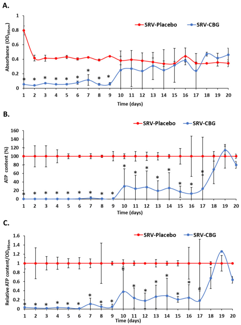
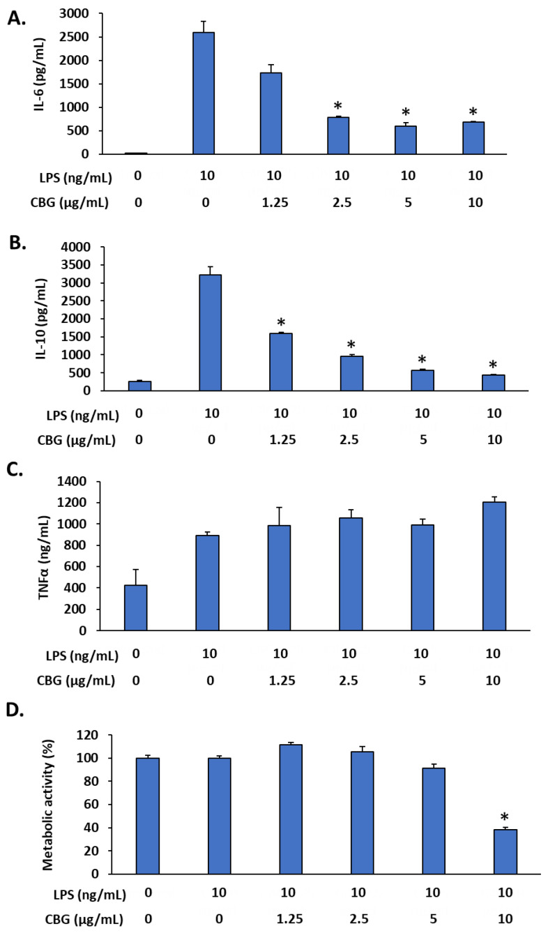
Synthetic surgical meshes are commonly used in abdominal wall reconstruction surgeries to strengthen a weak abdominal wall. Common mesh-related complications include local infection and inflammatory processes. Because cannabigerol (CBG) has both antibacterial and anti-inflammatory properties, we proposed that coating VICRYL (polyglactin 910) mesh with a sustained-release varnish (SRV) containing CBG would prevent these complications. We used an in vitro infection model with Staphylococcus aureus and an in vitro inflammation model of lipopolysaccharide (LPS)-stimulated macrophages. Meshes coated with either SRV-placebo or SRV-CBG were exposed daily to S. aureus in tryptic soy medium (TSB) or macrophage Dulbecco's modified eagle medium (DMEM). Bacterial growth and biofilm formation in the environment and on the meshes were assessed by changes in optical density, bacterial ATP content, metabolic activity, crystal violet staining, spinning disk confocal microscopy (SDCM), and high-resolution scanning electron microscopy (HR-SEM). The anti-inflammatory effect of the culture medium that was exposed daily to the coated meshes was analyzed by measuring the release of the cytokines IL-6 and IL-10 from LPS-stimulated RAW 264.7 macrophages with appropriate ELISA kits. Additionally, a cytotoxicity assay was performed on Vero epithelial cell lines. We observed that compared with SRV-placebo, the segments coated with SRV-CBG inhibited the bacterial growth of S. aureus in the mesh environment for 9 days by 86 ± 4% and prevented biofilm formation and metabolic activity in the surroundings for 9 days, with respective 70 ± 2% and 95 ± 0.2% reductions. The culture medium that was incubated with the SRV-CBG-coated mesh inhibited LPS-induced secretion of IL-6 and IL-10 from the RAW 264.7 macrophages for up to 6 days without affecting macrophage viability. A partial anti-inflammatory effect was also observed with SRV-placebo. The conditioned culture medium was not toxic to Vero epithelial cells, which had an IC50 of 25 µg/mL for CBG. In conclusion, our data indicate a potential role of coating VICRYL mesh with SRV-CBG in preventing infection and inflammation in the initial period after surgery.

Keywords: Staphylococcus aureus; biofilm; cannabigerol; cytokines; macrophages; mesh; sustained-release varnish.


Conflict of interest statement

The authors declare no conflict of interest.

Figures

Figure 1
Figure 1
HR-SEM images of SRV-placebo-coated meshes at ×100 (A,B) magnification. HR-SEM images of SRV-CBG-coated meshes at ×100 (C,D) magnification. HR-SEM images of uncoated meshes at ×100 (E,F) magnification.
Figure 2
Figure 2
Antibacterial effects of the SRV-CBG-coated mesh on the planktonic growth of S. aureus. (A). SRV-CBG- and SRV-placebo-coated meshes were incubated daily with fresh S. aureus cultures for 20 days, and the OD at 595 nm of the supernatant was measured daily after a 24 h incubation period. n = 3. * p < 0.05 for SRV-CBG versus SRV-placebo, according to Student’s t-test. (B). The percentage of ATP content in S. aureus planktonic bacteria following 24 h exposure to SRV-CBG-coated meshes in comparison to ATP contents in S. aureus planktonic bacteria exposed to SRV-placebo-coated meshes. The kinetic study was conducted for 20 days. n = 3. * p < 0.05 for SRV-CBG versus SRV-placebo, according to Student’s t-test. (C). The relative ATP content in comparison to cell density (OD595nm) for each sample. n = 3. * p < 0.05 for SRV-CBG versus SRV-placebo, according to Student’s t-test.
Figure 3
Figure 3
Antibacterial effects of SRV-CBG-coated mesh on planktonic growth of a clinical MDRSA CI-M isolate. SRV-CBG- and SRV-placebo-coated meshes were incubated daily with fresh MDRSA CI-M cultures for 6 days. The relative ATP content in MDRSA CI-M planktonic bacteria exposed to SRV-CBG-coated meshes was reduced by 78–99% compared to ATP contents in planktonic MDRSA CI-M bacteria exposed to SRV-placebo-coated meshes. The kinetic study was conducted for 6 days. n = 3. * p < 0.05 for SRV-CBG versus SRV-placebo, according to Student’s t-test.
Figure 4
Figure 4
(A). Biofilm mass of S. aureus formed on the plastic well after daily exposure to SRV-CBG-coated mesh or SRV-placebo-coated mesh, as determined by crystal violet staining. n = 3. * p < 0.05 for SRV-CBG versus SRV-placebo, according to Student’s t-test. (B). Biofilm metabolic activity of S. aureus exposed to SRV-CBG-coated mesh compared with biofilm metabolic activity of S. aureus exposed to SRV-placebo-coated mesh, as determined by using MTT assay. n = 3. * p < 0.05 for SRV-CBG versus SRV-placebo, according to Student’s t-test. (C). SYTO 9/PI staining of S. aureus biofilm formed on the plastic well after being exposed to SRV-placebo (upper row) and SRV-CBG-coated mesh (lower row) segments on day 5 of incubation.
Figure 5
Figure 5
Antibiofilm effects of SRV-CBG-coated meshes on a clinical MDRSA CI-M isolate. SRV-CBG- and SRV-placebo-coated meshes were incubated daily with fresh MDRSA CI-M cultures for 6 days. The metabolic activity of MDRSA CI-M biofilms formed following incubation with SRV-CBG-coated mesh was reduced by 50–95% compared with the metabolic activity of MDRSA CI-M biofilms formed following incubation with SRV-placebo-coated mesh, as determined by using MTT assay. n = 3. * p < 0.05 for SRV-CBG versus SRV-placebo, according to Student’s t-test.
Figure 6
Figure 6
HR-SEM images of biofilms formed on the plastic well 24 h after the fifth exposure of SRV-placebo (A,C,E) or SRV-CBG-coated (B,D,F) meshes to S. aureus. A ×5000 magnification is shown. Images from three different samples of each group are shown.
Figure 7
Figure 7
(AH) HR-SEM images of SRV-placebo (AD) and SRV-CBG-coated (EH) mesh surfaces after 5 days of incubation with S. aureus. Magnifications of ×2000 (A,B,E,F) and ×5000 (C,D,G,H) are shown.
Figure 8
Figure 8
Metabolic activity of the biofilms formed on the coated meshes, as determined by using the MTT assay. The metabolic activity of SRV-placebo-coated mesh was set to 100%. n = 3. * p < 0.05 for SRV-CBG versus SRV-placebo, according to Student’s t-test.
Figure 9
Figure 9
Biocompatibility assay on Vero epithelial cells. (A,B) Vero cell monolayer was exposed for 24 h to the daily collected DMEM-conditioned medium from SRV-CBG- and SRV-placebo-coated meshes for a period of 20 days. At the end of incubation, the cells were visualized under a light microscope and stained with CV for measuring the total cell mass (A) or exposed to MTT to measure the metabolic activity (B). n = 3. (C,D) Vero cell monolayer was incubated in the absence or presence of increasing concentrations of CBG (0–25 µg/mL) for 24 h and then either stained with CV for measuring their total cell mass (C) or MTT for measuring their metabolic activity (D). n = 3 * p < 0.05 in comparison to control (no CBG), according to Student’s t-test.
Figure 10
Figure 10
CBG inhibited LPS-induced IL-6 and IL-10 secretion but not TNFα secretion from RAW 264.7 macrophages. (AC) RAW 264.7 macrophages were exposed to increasing concentrations of CBG in the presence of LPS (10 ng/mL) for 24 h, and the IL-6 (A), IL-10 (B), and TNFα (C) levels were analyzed by using respective ELISA kits. (D) The metabolic activity of macrophages after a 6 h incubation period with various concentrations of CBG and/or LPS. n = 3; * p < 0.05 in comparison to LPS-treated cells, according to Student’s t-test.
Figure 11
Figure 11
SRV-CBG-coated meshes inhibited LPS-induced IL-6 and IL-10 secretion from RAW 264.7 macrophages. (A,B) The levels of IL-6 (A) and IL-10 (B) secreted by RAW 264.7 macrophages after exposure to the conditioned medium collected from SRV-placebo and SRV-CBG for 14 days and 10 ng/mL LPS. The black and green lines represent the amount of IL-6 and IL-10 secreted by the same macrophages in the presence of LPS (10 ng/mL) (black line) or LPS (10 ng/mL) with CBG (2.5 µg/mL) (green line), respectively. n = 3. * p < 0.05 in comparison to LPS-treated cells, according to Student’s t-test. (C) Metabolic activity of the RAW 264.7 macrophages exposed to the conditioned medium of SRV-CBG and SRV-placebo for 14 days, as determined by using the MTT assay. The black line shows the MTT values of untreated macrophages. n = 3.

Similar articles

Cited by

References

    1. Grafmiller K.T., Zuckerman S.T., Petro C., Liu L., von Recum H.A., Rosen M.J., Korley J.N. Antibiotic-releasing microspheres prevent mesh infection in vivo. J. Surg. Res. 2016;206:41–47. doi: 10.1016/j.jss.2016.06.099. - DOI - PMC - PubMed
    1. Arnold M.R., Kao A.M., Gbozah K.K., Heniford B.T., Augenstein V.A. Optimal management of mesh infection: Evidence and treatment options. Int. J. Abdom. Wall Hernia Surg. 2018;1:42.
    1. Corduas F., Lamprou D.A., Mancuso E. Next-generation surgical meshes for drug delivery and tissue engineering applications: Materials, design and emerging manufacturing technologies. Bio-Des. Manuf. 2021;4:278–310. doi: 10.1007/s42242-020-00108-1. - DOI
    1. Bakri M., Lovato F.C., Diosti G.d.M., Salles Y.L.S.d.G., Moreira P.H.B., Collaço L.M., Czeczko N.G., Malafaia O., Kubrusly L.F. Comparative analysis of tissular response after abdominal wall repair using polypropylene mesh and bovine pericardium mesh. Arq. Bras. Cir. Dig. 2022;34:e1527. doi: 10.1590/0102-672020200003e1527. - DOI - PMC - PubMed
    1. Wilson R.B., Farooque Y. Risks and prevention of surgical site infection after hernia mesh repair and the predictive utility of ACS-NSQIP. J. Gastrointest. Surg. 2022;26:950–964. doi: 10.1007/s11605-022-05248-6. - DOI - PMC - PubMed

Grants and funding

LinkOut - more resources