. 2023 May 19;16(5):769.
doi: 10.3390/ph16050769.

Canagliflozin Ameliorates Oxidative Stress and Autistic-like Features in Valproic-Acid-Induced Autism in Rats: Comparison with Aripiprazole Action

Affiliations

Canagliflozin Ameliorates Oxidative Stress and Autistic-like Features in Valproic-Acid-Induced Autism in Rats: Comparison with Aripiprazole Action

Mohammed Moutaz Nakhal et al. Pharmaceuticals (Basel). .

Abstract

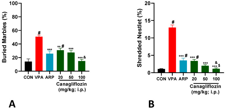
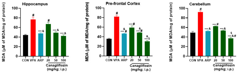
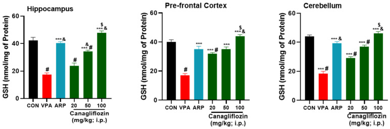
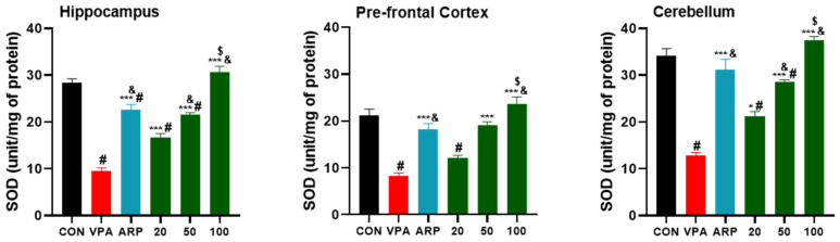
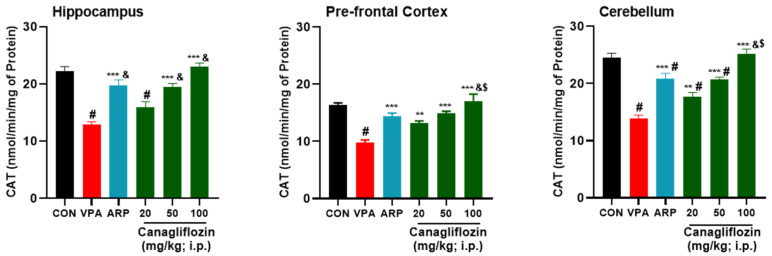
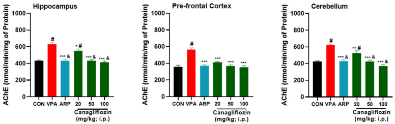
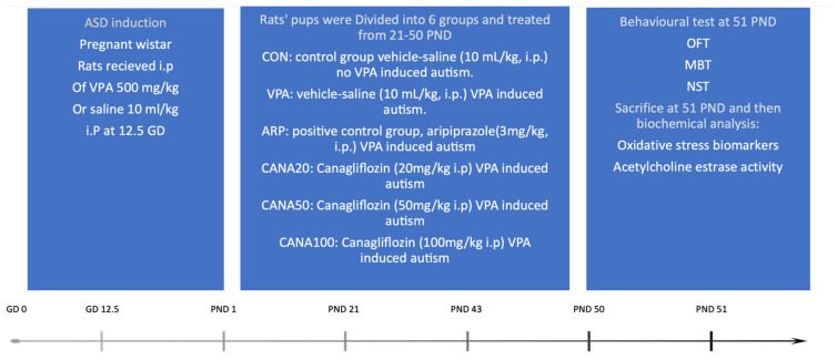
Based on their proven anti-inflammatory and antioxidant effects, recent studies have examined the therapeutic potential of the sodium-glucose cotransporter 2 (SGLT2) inhibitors in neurodevelopmental disorders such as autism spectrum disorder (ASD). Therefore, the aim of this study is to assess the effects of subchronic systemic treatment with intraperitoneal (i.p.) canagliflozin (20, 50, and 100 mg/kg) compared to aripiprazole (ARP) (3 mg/g, i.p.) in a valproic acid (VPA)-induced rat model of autism. The behavioral characteristics of ASD, oxidative stress, and acetylcholinesterase (AChE) activity in rats with ASD-like behaviors, which were induced by prenatal exposure to VPA, were evaluated. The behavioral assessment methods used for this study were the open field test (OFT), the marble-burying test (MBT), and the nestlet-shredding test (NST) to examine their exploratory, anxiety, and compulsiveness-like actions, while the biochemical assessment used for this study was an ELISA colorimetric assay to measure ASD biomarker activity in the hippocampus, prefrontal cortex, and cerebellum. Rats that were pretreated with 100 mg/kg of canagliflozin displayed a significantly lower percentage of shredding (1.12 ± 0.6%, p < 0.01) compared to the ARP group (3.52 ± 1.6%). Pretreatment with (20 mg/kg, 50 mg/kg, and 100 mg/kg) canagliflozin reversed anxiety levels and hyperactivity and reduced hyper-locomotor activity significantly (161 ± 34.9 s, p < 0.05; 154 ± 44.7 s, p < 0.05; 147 ± 33.6 s, p < 0.05) when compared with the VPA group (303 ± 140 s). Moreover, canagliflozin and ARP mitigated oxidative stress status by restoring levels of glutathione (GSH) and catalase (CAT) and increasing the levels of malondialdehyde (MDA) in all tested brain regions. The observed results propose repurposing of canagliflozin in the therapeutic management of ASD. However, further investigations are still required to verify the clinical relevance of canagliflozin in ASD.

Keywords: SGLT2 inhibitors; VPA-induced ASD; aripiprazole; autism spectrum disorder; behavioral assessments; biochemical assays; canagliflozin; oxidative stress biomarkers; rats.


Conflict of interest statement

The authors declare no financial or other types of competing interests with respect to the research, authorship, and/or publication of this article.

Figures

Figure 1
Figure 1
(A) Canagliflozin and aripiprazole mitigated stereotyped repetitive behavior in the marble-burying test (MBT). Marbles were counted after a 30 min testing session applying the same treatments. VPA-induced ASD rats treated with saline (VPA) displayed significantly increased repetitive behaviors when compared to control rats (CON) and all treatment groups. Canagliflozin (20, 50, and 100 mg/kg, i.p.) or the reference drug aripiprazole (ARP, 3 mg/kg, i.p.) was injected subchronically for 29 days in VPA-exposed rats. Values are expressed as mean ± SEM (n = 6). (B) Canagliflozin and aripiprazole attenuated increased obsessive–compulsive features in the nestlet-shredding test (NST). Repetitive nestlet-shredding behavior was measured after a 30 min testing session. VPA-exposed rats (VPA) demonstrated significantly elevated stereotyped repetitive behaviors compared to saline-exposed rats (CON). Canagliflozin (20, 50, and 100 mg/kg) or the reference drug aripiprazole (ARP, 3 mg/kg, i.p.) was administered subchronically for 29 days. The administration of aripiprazole (ARP, 3 mg/kg, i.p.) or canagliflozin (20, 50, or 100 mg/kg, i.p.) attenuated stereotyped repetitive behavior of VPA-exposed rats. Values are expressed as ± SEM (n = 6). # p < 0.05 when compared with the CON group, & p < 0.05 when compared with the canagliflozin 20 mg group, $ p < 0.05 when compared with the ARP group, ** p < 0.01 when compared with the VPA group, *** p < 0.001 when compared with the VPA group.
Figure 2
Figure 2
Canagliflozin and aripiprazole decrease oxidative stress biomarker (MDA) in the hippocampus, prefrontal cortex, and cerebellum. # p < 0.05 when compared with the CON group, & p < 0.05 when compared with the canagliflozin 20 mg group, $ p < 0.05 when compared with the ARP group, *** p < 0.001 when compared with the VPA group. Values are expressed as mean ± SEM (n = 6).
Figure 3
Figure 3
Canagliflozin and aripiprazole increase antioxidant (GSH) in the hippocampus, prefrontal cortex, and cerebellum. # p < 0.05 when compared with the CON group, & p < 0.05 when compared with the canagliflozin 20 mg group, $ p < 0.05 when compared with the ARP group, *** p < 0.001 when compared with the VPA group. Values are expressed as mean ± SEM (n = 6).
Figure 4
Figure 4
Canagliflozin and aripiprazole elevate antioxidant (SOD) in the hippocampus, prefrontal cortex, and cerebellum. # p < 0.05 when compared with the CON group, & p < 0.05 when compared with the canagliflozin 20 mg group, $ p < 0.05 when compared with the ARP group * p < 0.05 when compared with VPA group. *** p < 0.001 when compared with the VPA group. Values are expressed as mean ± SEM (n = 6).
Figure 5
Figure 5
Canagliflozin and aripiprazole reduced neurotoxicity biomarker (AChE) levels in the hippocampus, prefrontal cortex, and cerebellum of VPA-exposed rats. # p < 0.05 when compared with the CON group, & p < 0.05 when compared with the canagliflozin 20 mg group, $ p < 0.05 when compared with the ARP group, ** p < 0.01 when compared with the VPA group, *** p < 0.001 when compared with the VPA group. Values are expressed as mean ± SEM (n = 6).
Figure 6
Figure 6
Canagliflozin and aripiprazole enhance antioxidant (CAT) in the hippocampus, prefrontal cortex, and cerebellum. # p < 0.05 when compared with the CON group, & p < 0.05 when compared with the canagliflozin 20 mg group, * p < 0.05 when compared with the VPA group, ** p < 0.01 when compared with the VPA group, *** p < 0.001 when compared with the VPA group. Values are expressed as mean ± SEM (n = 6).
Figure 7
Figure 7
Graphical illustration of the implemented project plan, treatments, behavioral examinations, and biochemical analysis with autistic and control Wistar rats. At gestational day 12.5 (GD 12.5), pregnant rats were injected intraperitonially either with VPA (500 mg/kg) or saline (10 mL/kg). After delivery, starting from postnatal day 21, male and female pups were subdivided into 6 groups (6 rats/group) and received intraperitoneally (i.p.) the following treatments until PND 50, which is shown in the experimental design. Group 1: control rats injected with saline (10 mL/kg). Group 2: VPA-induced autism rats were injected with saline (10 mL/kg). Group 3: VPA-induced autism rats were injected with aripiprazole (3 mg/kg) and considered as the positive control. Group 4: VPA-induced autism rats were injected with 20 mg/kg of canagliflozin. Group 5: VPA-induced autistic rats were injected with 50 mg/kg of canagliflozin. Group 6: VPA-exposed autistic rats were injected with 100 mg/kg of canagliflozin. All 6 groups were subjected to behavioral assessment at PND 51 and scarified to undergo biochemical assays. OFT—open field test; MBT—marble-burying test; NST—nestlet-shredding test.

Similar articles

Cited by

References

    1. Jones E.J.H., Gliga T., Bedford R., Charman T., Johnson M.H. Developmental pathways to autism: A review of prospective studies of infants at risk. Neurosci. Biobehav. Rev. 2014;39:1–33. doi: 10.1016/j.neubiorev.2013.12.001. - DOI - PMC - PubMed
    1. Nicholas J.S., Charles J.M., Carpenter L.A., King L.B., Jenner W., Spratt E.G. Prevalence and characteristics of children with autism-spectrum disorders. Ann. Epidemiol. 2008;18:130–136. doi: 10.1016/j.annepidem.2007.10.013. - DOI - PubMed
    1. Medavarapu S., Marella L.L., Sangem A., Kairam R. Where is the evidence? A narrative literature review of the treatment modalities for autism spectrum disorders. Cureus. 2019;11:e3901. doi: 10.7759/cureus.3901. - DOI - PMC - PubMed
    1. Varamini B., Sikalidis A.K., Bradford K.L. Resveratrol increases cerebral glycogen synthase kinase phosphorylation as well as protein levels of drebrin and transthyretin in mice: An exploratory study. Int. J. Food Sci. Nutr. 2014;65:89–96. doi: 10.3109/09637486.2013.832171. - DOI - PubMed
    1. Indika N.L., Frye R.E., Rossignol D.A., Owens S.C., Senarathne U.D., Grabrucker A.M., Perera R., Engelen M.P., Deutz N.E. The Rationale for Vitamin, Mineral, and Cofactor Treatment in the Precision Medical Care of Autism Spectrum Disorder. J. Pers. Med. 2023;13:252. doi: 10.3390/jpm13020252. - DOI - PMC - PubMed

LinkOut - more resources