. 2023 Apr 26;15(5):1340.
doi: 10.3390/pharmaceutics15051340.

Microporous/Macroporous Polycaprolactone Scaffolds for Dental Applications

Affiliations

Microporous/Macroporous Polycaprolactone Scaffolds for Dental Applications

Tara Shabab et al. Pharmaceutics. .

Abstract

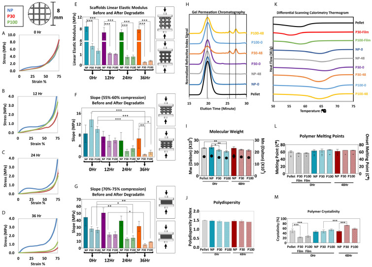
This study leverages the advantages of two fabrication techniques, namely, melt-extrusion-based 3D printing and porogen leaching, to develop multiphasic scaffolds with controllable properties essential for scaffold-guided dental tissue regeneration. Polycaprolactone-salt composites are 3D-printed and salt microparticles within the scaffold struts are leached out, revealing a network of microporosity. Extensive characterization confirms that multiscale scaffolds are highly tuneable in terms of their mechanical properties, degradation kinetics, and surface morphology. It can be seen that the surface roughness of the polycaprolactone scaffolds (9.41 ± 3.01 µm) increases with porogen leaching and the use of larger porogens lead to higher roughness values, reaching 28.75 ± 7.48 µm. Multiscale scaffolds exhibit improved attachment and proliferation of 3T3 fibroblast cells as well as extracellular matrix production, compared with their single-scale counterparts (an approximate 1.5- to 2-fold increase in cellular viability and metabolic activity), suggesting that these structures could potentially lead to improved tissue regeneration due to their favourable and reproducible surface morphology. Finally, various scaffolds designed as a drug delivery device were explored by loading them with the antibiotic drug cefazolin. These studies show that by using a multiphasic scaffold design, a sustained drug release profile can be achieved. The combined results strongly support the further development of these scaffolds for dental tissue regeneration applications.

Keywords: architecture; biomateriomics; biomimetic; dental scaffolds; drug delivery; multiphasic.


Conflict of interest statement

The authors declare no conflict of interest. The funders had no role in the design of the study; in the collection, analyses, or interpretation of data; in the writing of the manuscript; or in the decision to publish the results.

Figures

Figure 1
Figure 1
The workflow used in this study, showing the steps involved in manufacturing, scaffold characterisation, in vitro biological response and in vitro drug delivery.
Figure 2
Figure 2
(A) Schematic presentation of the manufacturing of PCL multiphasic scaffolds. (B) SEM of the porogen particles, (a,b) different magnifications of P30 porogen, (c,d) different magnifications of P100 porogen. (C) Porogen size distribution determined by dynamic light scattering (n = 5). (D) TGA analysis showing the decomposition temperature of the materials. (E) Rheological properties of the materials (PCL pellets, P30, and P100 films) (n = 3).
Figure 3
Figure 3
(A) Composite leaching process and SEM micrographs. (B) X-ray diffraction analysis of the scaffolds, confirming salt leaching (n = 3).
Figure 4
Figure 4
Mechanical compression testing performed in air (A) and in PBS (B) (n = 6); (C) Elastic modulus of the scaffolds; (D) Slope of the curve 55–60% compression; (E) Slope of the curve 70–75% compression; (F) Stereomicroscope imaging; (G) the micro-CT 3D reconstruction shows the general topography of the scaffolds; (H) SEM and (I) 3D profiles show the topography of the scaffolds; (J) Scaffold surface roughness (n = 9). Data are presented as mean ± SD. * and *** indicate p < 0.033 and p < 0.001, respectively.
Figure 5
Figure 5
(AG) Mechanical compression test during degradation (n = 5). (HJ) GPC shows molecular weight reduction and an increase in polydispersity (n = 3). (KM) DSC shows the change in crystallinity during 48 h of accelerated degradation (n = 3). Data are presented as mean ± SD. *, ** and *** in figures indicate p < 0.033, p < 0.002 and p < 0.001, respectively.
Figure 6
Figure 6
(A) Scanning electron microscopy of nonporous scaffolds during degradation. SEM micrographs of P30 (B) and P100 (C). (D) micro-CT and (E) mercury intrusion porosimetry (n = 3) show pore interconnectivity of the porous scaffolds (P30 and P100). (F) micro-CT 2D and 3D porosimetry before and after degradation (n = 5). (G) microporosity 3D visualization and pore size distribution/pore equivalent diameter (µm) of the scaffolds by micro-CT (n = 5). *, ** and *** in figures indicate p < 0.033, p < 0.002 and p < 0.001, respectively.
Figure 7
Figure 7
(A) PrestoBlue cell metabolic activity. Data showed an increase in cell metabolic activity from Day 3 to Day 21; data are presented as mean ± SD. *, ** and *** indicate p < 0.033, p < 0.002 and p < 0.001 respectively. (B) SEM images of cultured fibroblast cells after 3 and 10 days of culture. (C) Representative confocal microscopy images of fibroblast cells cultured on NP, P30 and P100 scaffolds at Day 3 and Day 10. Cells were labelled with antibodies to visualize Nuclei (DAPI; blue), FAK (green) and F-actin (Phalloidin; red).
Figure 8
Figure 8
(A) SEM of PCL films showing the surface topography before the experiment. (B) Water contact angle (wettability) of PCL films (n = 4). (C) Whole blood attachment and whole blood clotting on PCL films. (D) Fibre diameter, measured from SEM micrographs, (n = 6). Data are presented as mean ± SD. *, ** and *** in figures indicate p < 0.033, p < 0.002 and p < 0.001, respectively.
Figure 9
Figure 9
(A) Drug release profile. Data are presented as mean ± SD (n = 8). (B) Antibacterial inhibition zone. Positive control (Cont+) is OxoidTM cefazolin antimicrobial susceptibility disks (30 µg); Negative control (Cont−) is drug-free scaffolds. Data are presented as mean ± SD (n = 3).

Similar articles

Cited by

References

    1. Mohtashami Z., Esmaili Z., Vakilinezhad M., Seyedjafari E., Javar H.A., Seyedjafari S.E. Pharmaceutical implants: Classification, limitations and therapeutic applications. Pharm. Dev. Technol. 2019;25:116–132. doi: 10.1080/10837450.2019.1682607. - DOI - PubMed
    1. Tesfamariam B. Bioresorbable vascular scaffolds: Biodegradation, drug delivery and vascular remodeling. Pharmacol. Res. 2016;107:163–171. doi: 10.1016/j.phrs.2016.03.020. - DOI - PubMed
    1. Dorati R., DeTrizio A., Modena T., Conti B., Benazzo F., Gastaldi G., Genta I. Biodegradable Scaffolds for Bone Regeneration Combined with Drug-Delivery Systems in Osteomyelitis Therapy. Pharmaceuticals. 2017;10:96. doi: 10.3390/ph10040096. - DOI - PMC - PubMed
    1. Guarino V., Ambrosio L. Temperature-driven processing techniques for manufacturing fully interconnected porous scaffolds in bone tissue engineering. Proc. Inst. Mech. Eng. Part H J. Eng. Med. 2010;224:1389–1400. doi: 10.1243/09544119JEIM744. - DOI - PubMed
    1. Bertassoni L.E.C., editor. Engineering Mineralized and Load Bearing Tissues, Advances in Experimental Medicine and Biology. Volume 881 Springer International Publishing; Cham, Switzerland: 2015. - PubMed

LinkOut - more resources