. 2023 Apr 28;15(5):1350.
doi: 10.3390/pharmaceutics15051350.

Saponin Fraction CIL1 from Lysimachia ciliata L. Enhances the Effect of a Targeted Toxin on Cancer Cells

Affiliations

Saponin Fraction CIL1 from Lysimachia ciliata L. Enhances the Effect of a Targeted Toxin on Cancer Cells

Paulina Koczurkiewicz-Adamczyk et al. Pharmaceutics. .

Abstract

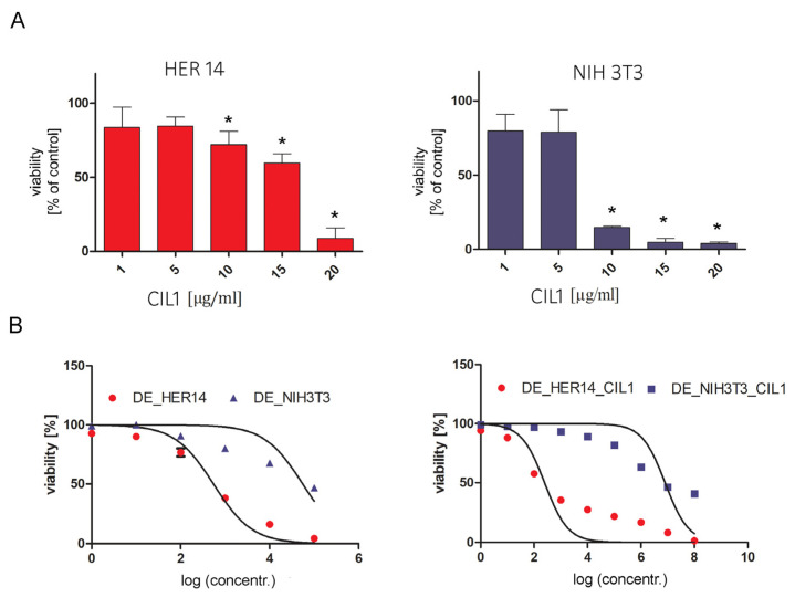
Saponins are plant metabolites that possess multidirectional biological activities, among these is antitumor potential. The mechanisms of anticancer activity of saponins are very complex and depend on various factors, including the chemical structure of saponins and the type of cell they target. The ability of saponins to enhance the efficacy of various chemotherapeutics has opened new perspectives for using them in combined anticancer chemotherapy. Co-administration of saponins with targeted toxins makes it possible to reduce the dose of the toxin and thus limit the side effects of overall therapy by mediating endosomal escape. Our study indicates that the saponin fraction CIL1 of Lysimachia ciliata L. can improve the efficacy of the EGFR-targeted toxin dianthin (DE). We investigated the effect of cotreatment with CIL1 + DE on cell viability in a 3-(4,5-dimethylthiazol-2-yl)-2,5-diphenyltetrazolium bromide (MTT) assay, on proliferation in a crystal violet assay (CV) and on pro-apoptotic activity using Annexin V/7 Actinomycin D (7-AAD) staining and luminescence detection of caspase levels. Cotreatment with CIL1 + DE enhanced the target cell-specific cytotoxicity, as well as the antiproliferative and proapoptotic properties. We found a 2200-fold increase in both the cytotoxic and antiproliferative efficacy of CIL1 + DE against HER14-targeted cells, while the effect on control NIH3T3 off-target cells was less profound (6.9- or 5.4-fold, respectively). Furthermore, we demonstrated that the CIL1 saponin fraction has a satisfactory in vitro safety profile with a lack of cytotoxic and mutagenic potential.

Keywords: CIL1 saponin fraction; anti-cancer activity; endosomal escape; ribosome-inactivating protein; targeted therapy; targeted toxin.


Conflict of interest statement

The authors declare no conflict of interest.

Figures

Figure 1
Figure 1
Influence of CIL 1 on the combined induction cytotoxicity by DE in HER14 and NIH3T3 cells. The non-toxic concentration of CIL1 (3 µg/mL) confirmed in the MTT assay (A) was used in the study. HER14 or NIH 3T3 cells were seeded at an initial density of 2 × 10³ cells/well in 96-well plates. Cells were incubated for 72 h with DE in the presence or absence of CIL1. Cell viability was determined using an MTT assay (B). Each experiment was repeated in triplicate. Statistical significance (*) was calculated relative to control (p < 0.05) using Brown Forsythe and Welsch ANOVA test, along with a post-hoc unpaired t-test with Welch’s correction.
Figure 2
Figure 2
Influence of CIL 1 on the combined induction of DE proliferation inhibition in HER14 and NIH3T3 cells. HER14 or NIH 3T3 cells were seeded at an initial density of 2 × 10³ cells/well in 96-well plates. Cells were incubated for 72 h with DE in the presence or absence of CIL1 (3 µg/mL). Cell proliferation was measured using a crystal violet assay. Each experiment was repeated in triplicate.
Figure 3
Figure 3
Influence of CIL 1 on the combined induction of apoptosis by DE in HER14 and NIH3T3 cells. Cells were incubated for 48 h with DE in the presence or absence of CIL1 (3 µg/mL) (flow cytometry analysis) or 24 h for luminescence analysis. Cell apoptosis was detected using (A) Annexin V/7-AAD staining followed by flow cytometry analysis and (B) luminescent detection of caspase 3/7. Each experiment was repeated in triplicate. Statistical significance (*) was calculated relative to control (p < 0.05) using Brown Forsythe and Welsch ANOVA test, along with a post-hoc unpaired t-test with Welch’s correction.
Figure 4
Figure 4
Cytotoxicity (A) and mutagenicity (B) of CIL1. The neurotoxicity, hepatotoxicity and cardiotoxicity of CIL1 were evaluated using the following cell lines: human neuroblastoma (SHSY-5Y), human hepatocellular carcinoma (HepG2) and rat cardiomyoblast (H9c2). Cells were incubated in the presence of CIL1 in the concentration range of 1–20 µg/mL for 72 h, then MTT and CytoTox Glo assays were performed. The graphs represent the mean percent of viability/cytotoxicity ± SD compared to untreated cells (control) (A). Each experiment was repeated in triplicate. The mutagenicity was evaluated using the Ames test. Salmonella typhimurium strains TA98 and TA100 were used in the study in the absence (−S9) or presence (+S9) of the S9 fraction. Bacterial cultures were incubated for 48 h with CIL1 (10 µg/mL) or positive controls 2-nitrofluorene (NF-2) at 2.0 μg/mL and 4-nitroquinoline-1-oxide (NQNO4) at 0.1 μg/mL (B). Each bar represents the average number of revertants/plate (± SD). Statistical significance (*) was calculated relative to control (p < 0.05) using Brown Forsythe and Welsch ANOVA test, along with a post-hoc unpaired t-test with Welch’s correction.

Similar articles

Cited by

References

    1. Magedans Y.V., Yendo A.C., Costa F., Gosmann G., Fett-Neto A.G. Foamy matters: An update on Quillaja saponins and their use as immunoadjuvants. Future Med. Chem. 2019;11:1485–1499. doi: 10.4155/fmc-2018-0438. - DOI - PubMed
    1. Wang P. Natural and Synthetic Saponins as Vaccine Adjuvants. Vaccines. 2021;9:222. doi: 10.3390/vaccines9030222. - DOI - PMC - PubMed
    1. Koczurkiewicz P., Kowolik E., Podolak I., Wnuk D., Piska K., Łabędź-Masłowska A., Wójcik-Pszczoła K., Pękala E., Czyż J., Michalik M. Synergistic Cytotoxic and Anti-invasive Effects of Mitoxantrone and Triterpene Saponins from Lysimachia ciliata on Human Prostate Cancer Cells. Planta Med. 2016;82:1546–1552. doi: 10.1055/s-0042-117537. - DOI - PubMed
    1. Wang B., Min W., Lin S., Song L., Yang P., Ma Q., Guo J. Saikosaponin-d increases radiation-induced apoptosis of hepatoma cells by promoting autophagy via inhibiting mTOR phosphorylation. Int. J. Med. Sci. 2021;18:1465–1473. doi: 10.7150/ijms.53024. - DOI - PMC - PubMed
    1. Wang K., Zhan Y., Chen B., Lu Y., Yin T., Zhou S., Zhang W., Liu X., Du B., Wei X., et al. Tubeimoside I-induced lung cancer cell death and the underlying crosstalk between lysosomes and mitochondria. Cell Death Dis. 2020;11:708. doi: 10.1038/s41419-020-02915-x. - DOI - PMC - PubMed