. 2023 May 13;15(5):1489.
doi: 10.3390/pharmaceutics15051489.

Mesenchymal Stromal Cell Exosomes Mediate M2-like Macrophage Polarization through CD73/Ecto-5'-Nucleotidase Activity

Affiliations

Mesenchymal Stromal Cell Exosomes Mediate M2-like Macrophage Polarization through CD73/Ecto-5'-Nucleotidase Activity

Kristeen Ye Wen Teo et al. Pharmaceutics. .

Abstract

Mesenchymal stem/stromal cell (MSC) exosomes have been shown to alleviate immune dysfunction and inflammation in preclinical animal models. This therapeutic effect is attributed, in part, to their ability to promote the polarization of anti-inflammatory M2-like macrophages. One polarization mechanism has been shown to involve the activation of the MyD88-mediated toll-like receptor (TLR) signaling pathway by the presence of extra domain A-fibronectin (EDA-FN) within the MSC exosomes. Here, we uncovered an additional mechanism where MSC exosomes mediate M2-like macrophage polarization through exosomal CD73 activity. Specifically, we observed that polarization of M2-like macrophages by MSC exosomes was abolished in the presence of inhibitors of CD73 activity, adenosine receptors A2A and A2B, and AKT/ERK phosphorylation. These findings suggest that MSC exosomes promote M2-like macrophage polarization by catalyzing the production of adenosine, which then binds to adenosine receptors A2A and A2B to activate AKT/ERK-dependent signaling pathways. Thus, CD73 represents an additional critical attribute of MSC exosomes in mediating M2-like macrophage polarization. These findings have implications for predicting the immunomodulatory potency of MSC exosome preparations.

Keywords: CD73; exosomes; extracellular vesicles; immunomodulation; macrophage; mesenchymal stromal/stem cells.


Conflict of interest statement

The authors report that they have no conflict of interest in this study. S.K.L. holds founder shares in Paracrine Therapeutics Pte Ltd.

Figures

Figure 1
Figure 1
Time schedule for treatment of primary macrophages with MSC exosomes and inhibitors in vitro. (A) Primary macrophages were treated with MSC exosomes and harvested at 24 and 48 h for qPCR and IF. (B) Primary macrophages were co-treated with MSC exosomes and PSB12379 (CD73 inhibitor), before being harvested at 30 min for WB, and at 24 and 48 h for qPCR and IF analyses. (C) Primary macrophages were pre-treated with ZM241385 (A2A receptor inhibitor), PSB1115 (A2B receptor inhibitor), wortmannin (AKT inhibitor) or U0126 (ERK inhibitor) prior to exosome treatment, before being harvested at 30 min for WB, and at 24 and 48 h for qPCR and IF analyses. WB, western blot; qPCR, quantitative reverse transcription polymerase chain reaction; IF, immunofluorescence.
Figure 2
Figure 2
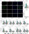
MSC exosomes promote M2-like but not M1-like macrophage polarization. (A) IF staining for CD206 and CD68 at 48 h. Representative images (n = 3). Scale bar: 100 μm. (B) Quantitative MFI of CD206 and CD68 at 48 h. (C) Gene expression analysis of M2-associated genes at 24 and 48 h. (D) IF staining for iNOS and CD68 at 48 h. Representative images (n = 3). Scale bar: 100 μm. (E) Quantitative MFI of iNOS and CD68 at 48 h. (F) Gene expression analysis of M1-associated genes at 24 and 48 h. Data are presented as mean ± SD. * p < 0.05, ** p < 0.01, and *** p < 0.001 compared to M0 macrophages. n = 3/group.
Figure 3
Figure 3
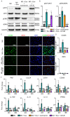
Exosomal CD73 mediates the effects of MSC exosomes in M2-like macrophage polarization. (A) Presence of CD73 protein band at ~70 kDa. (B) Percentage of CD73 activity in MSC exosomes following treatment with CD73 inhibitor (PSB12379) for 60 min. (C) IF staining of CD206 and iNOS in primary macrophages treated with MSC exosomes in presence or absence of CD73 inhibition (PSB12379) for 48 h. Representative images (n = 3). Scale bar: 100 μm. Quantitative MFI of (D) CD206 and (E) iNOS at 48 h. (F) Gene expression analysis of M2-associated genes at 24 and 48 h. Data are presented as mean ± SD. * p < 0.05, ** p < 0.01, *** p < 0.001 compared to M0, # p < 0.05, ## p < 0.01, and ### p < 0.001 compared to Exo or M0 + Exo group. n = 3–4/group.
Figure 4
Figure 4
Exosomal CD73 mediates activation of AKT and ERK signaling through specific adenosine receptors A2A and A2B. (A) Western blotting and (B) semi-quantitative analysis of AKT and ERK phosphorylation in primary macrophages treated with MSC exosomes in the presence or absence of CD73 inhibition by PSB12379. Representative images (n = 3). (C) Western blotting and (D) semi-quantitative analysis of AKT and ERK phosphorylation in primary macrophages pre-treated with A2A receptor inhibitor (ZM241385) and A2B receptor inhibitor (PSB1115) before exosome treatment. Representative images (n = 3). Data are presented as mean ± SD. * p < 0.05, ** p < 0.01, *** p < 0.001 compared to M0, # p < 0.05, ## p < 0.01, and ###p < 0.001 compared to M0 + Exo. n = 3–4/group.
Figure 5
Figure 5
MSC exosomes modulate macrophage polarization through specific adenosine receptors A2A and A2B. (A) IF staining at 48 h for CD206 and iNOS in primary macrophages treated with MSC exosomes, following pre-treatment with adenosine A2A receptor inhibitor (ZM241385) and A2B receptor inhibitor (PSB1115). Representative images (n = 3). Scale bar: 100 μm. Quantitative MFI of (B) CD206 and (C) iNOS at 48 h. (D) Gene expression analysis of M2-associated genes at 24 and 48 h. Data are presented as mean ± SD. * p < 0.05, ** p < 0.01, *** p < 0.001 compared to M0, # p < 0.05, ## p < 0.01, and ### p < 0.001 compared to M0 + Exo. n = 3–4/group.
Figure 6
Figure 6
MSC exosomes promote M2 macrophage polarization via AKT and ERK signaling pathways. (A) Western blotting and (B) semi-quantitative analysis of AKT and ERK phosphorylation in primary macrophages pre-treated with AKT inhibitor (wortmannin) or ERK inhibitor (U0126) before exosome treatment. Representative images (n = 3). (C) IF staining at 48 h for CD206 and iNOS in primary macrophages treated with MSC exosomes, following pre-treatment with AKT inhibitor (wortmannin) and ERK inhibitor (U0126). Representative images (n = 3). Scale bar: 100 μm. Quantitative MFI of (D) CD206 and (E) iNOS at 48 h. (F) Gene expression analysis of M2-associated genes at 24 and 48 h. Data are presented as mean ± SD. * p < 0.05, ** p < 0.01, *** p < 0.001 compared to M0, # p < 0.05, ## p < 0.01, and ### p < 0.001 compared to M0 + Exo. n = 3–4/group.
Figure 7
Figure 7
Proposed mechanism of MSC exosomes in promoting M2 macrophage polarization. Exosomal CD73 converts AMP to adenosine, which in turn interacts with A2A receptor to activate AKT and ERK signaling, and with A2B receptor to activate AKT signaling to promote M2 macrophage polarization. Image is created with Biorender.com on 8th May 2023.

References

    1. Toh W.S., Zhang B., Lai R.C., Lim S.K. Immune regulatory targets of mesenchymal stromal cell exosomes/small extracellular vesicles in tissue regeneration. Cytotherapy. 2018;20:1419–1426. doi: 10.1016/j.jcyt.2018.09.008. - DOI - PubMed
    1. Watanabe S., Alexander M., Misharin A.V., Budinger G.R.S. The role of macrophages in the resolution of inflammation. J. Clin. Invest. 2019;129:2619–2628. doi: 10.1172/JCI124615. - DOI - PMC - PubMed
    1. Mills C. M1 and M2 macrophages: Oracles of health and disease. Crit. Rev. ™ Immunol. 2012;32:463–488. doi: 10.1615/CritRevImmunol.v32.i6.10. - DOI - PubMed
    1. Okamoto T., Gohil K., Finkelstein E.I., Bove P., Akaike T., van der Vliet A. Multiple contributing roles for NOS2 in LPS-induced acute airway inflammation in mice. Am. J. Physiol. Lung Cell Mol. Physiol. 2004;286:L198–L209. doi: 10.1152/ajplung.00136.2003. - DOI - PubMed
    1. Zeidler P.C., Millecchia L.M., Castranova V. Role of inducible nitric oxide synthase-derived nitric oxide in lipopolysaccharide plus interferon-γ-induced pulmonary inflammation. Toxicol. Appl. Pharmacol. 2004;195:45–54. doi: 10.1016/j.taap.2003.10.005. - DOI - PubMed