. 2023 May 18;15(5):1527.
doi: 10.3390/pharmaceutics15051527.

Lung Inflammation Resolution by RvD1 and RvD2 in a Receptor-Dependent Manner

Affiliations

Lung Inflammation Resolution by RvD1 and RvD2 in a Receptor-Dependent Manner

Jin Gao et al. Pharmaceutics. .

Abstract

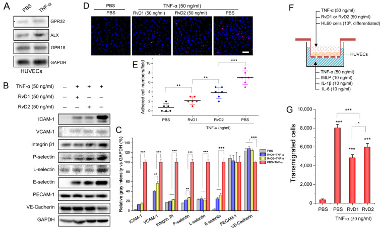
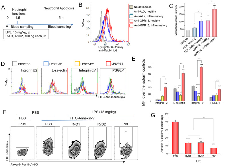
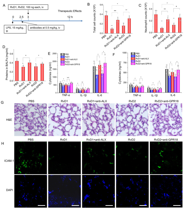
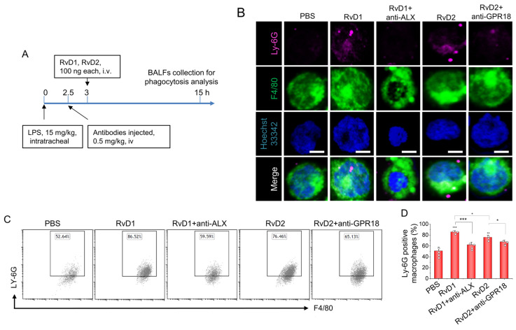
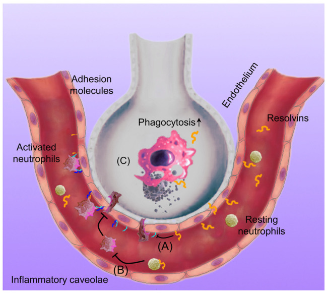
Inflammation resolution is an active process via specialized pro-resolving mediators (SPMs) to fight invading microbes and repair tissue injury. RvD1 and RvD2 are SPMs produced from DHA during inflammation responses and show a benefit in treating inflammation disorders, but it is not completely understood how they act on vasculature and immune cells in the lung to promote inflammation resolution programs. Here, we studied how RvD1 and RvD2 regulated the interactions between endothelial cells and neutrophils in vitro and in vivo. In an acute lung inflammation (ALI) mouse model, we found that RvD1 and RvD2 resolved lung inflammation via their receptors (ALX/GPR32 or GPR18) and enhanced the macrophage phagocytosis of apoptotic neutrophils, which may be the molecular mechanism of lung inflammation resolution. Interestingly, we observed the higher potency of RvD1 over RvD2, which may be associated with unique downstream signaling pathways. Together, our studies suggest that the targeted delivery of these SPMs into inflammatory sites may be novel strategies with which to treat a wide range of inflammatory diseases.

Keywords: ALX/GPR32; GPR18; RvD1; RvD2; acute lung inflammation (ALI); inflammation resolution; neutrophils.


Conflict of interest statement

The authors declare no conflict of interest.

Figures

Figure 1
Figure 1
RvD1 and RvD2 regulate the expression of adhesion molecules in HUVECs and inhibit leukocyte adherence as well as transmigration in vitro. (A) The receptors of RvD1 and RvD2 expressed on HUVECs 4 h after being treated with 50 ng/mL TNF-α. (B) RvD1 and RvD2 downregulate adhesion molecules of HUVECs, and their quantification (C). (D) Confocal images show the adhesion of HL-60 to HUVECs after treatment with RvD1 or RvD2. A 100% confluence of HUVECs were treated with 50 ng/mL TNF-α for 4 h and incubated with 105 neutrophil-like HL60 cells fluorescently labeled with DiD. (E) The quantification of confocal images in (D). (F) Transwell assay to measure the transmigration of neutrophils (HL60 cells) across confluent HUVECs and (G) the quantification results of (F). The data are expressed as means ± SD, n = 3–6. A one-way ANOVA was applied to determine the difference among multiple groups with * p ≤ 0.05, ** p ≤ 0.01, and *** p ≤ 0.001.
Figure 2
Figure 2
RvD1 and RvD2 attenuate the expression of neutrophil adhesion molecules and increase neutrophil apoptosis in vivo. (A) The animal protocol for analyzing neutrophil functions after the treatment with RvD1 and RvD2. (B) The expression of RvD1 and RvD2 receptors in mouse neutrophils via flow cytometry, and their quantitative analysis (C). (D) Adhesion molecules on neutrophils regulated by RvD1 and RvD2, and their quantification (E). (F) RvD1 and RvD2 promote the apoptosis of inflammatory neutrophils in mouse blood 5 h after LPS challenge of a 15 mg/kg i.p. injection, and their quantitative analysis (G). The data are expressed as the mean ± SDs, n = 3. A one-way ANOVA was applied to determine the difference among multiple groups, with * p ≤ 0.05, ** p ≤ 0.01, and *** p ≤ 0.001.
Figure 3
Figure 3
Lung inflammation resolution is dependent on RvD1 or RvD2 action on their receptors expressed on neutrophils and the endothelium in an acute lung inflammation model. (A) The animal experiment protocol with which to address therapeutic effects of RvD1 or RvD2. The animals were challenged with 15 mg/kg LPS and, 3 h later, the animals were treated with resolvins with or without the corresponding receptor antibodies. The animal blood, lung tissues, and BALFs were collected to measure inflammatory responses. Total cells (B) and neutrophils (C) in BALFs. (D) Protein contents in BALFs were determined via the BCA method. Cytokines, including TNF-α, IL-1β, and IL-6, in plasma (E) and BALFs (F) were determined by ELISA kits. (G) Typical H&E staining images of the lungs in each group. Magnification 100×. (H) Immunofluorescent images of ICAM-1 expression in the lung. Scale bar 100 µm. The data are expressed as the mean ± SDs, n = 3. A one-way ANOVA was applied to determine the difference among multiple groups, with * p ≤ 0.05, ** p ≤ 0.01, and *** p ≤ 0.001.
Figure 4
Figure 4
Phagocytosis of apoptotic neutrophils by macrophages is a mechanism of lung inflammation resolution in acute lung inflammation (ALI) induced by LPS. (A) The animal experiment protocol with which to address macrophage phagocytosis in the lungs. (B) At a time of 12 h after treatment with resolvins in ALI, macrophages in BALFs were collected and fixed. The anti-LY-6G antibody was used to identify neutrophils or related apoptotic bodies after macrophage membranes were permeabilized. The colocalization of LY-6G and F4/80 signals indicates the phagocytosis of apoptotic neutrophils by macrophages. Scale bar 10 µm. (C) To quantitatively analyze phagocytosis, flow cytometry was conducted to measure percentages of neutrophils or apoptotic bodies containing macrophages, and their quantitative analysis (D). The data are expressed as the mean ± SDs, n = 3. A one-way ANOVA was applied to determine the difference among multiple groups, with * p ≤ 0.05, ** p ≤ 0.01, and *** p ≤ 0.001.
Figure 5
Figure 5
The molecular mechanism of RvD1/RvD2 in regulating the inflammation resolution in the lung. LPS induces an inflammatory response in the lungs, and endothelial cells as well as neutrophils upregulate their adhesion molecules to promote neutrophil adhesion and transmigration in the lungs. If the inflammation is out of control, it may cause tissue damage and chronic inflammation; however, when treating it with resolvins, the adhesion molecule expression on endothelial cells (A) and neutrophils (B) is alleviated, thus mitigating neutrophil transmigration and neutrophil apoptosis. Next, resolvins enhance the macrophage phagocytosis of apoptotic neutrophils (C), thus maintaining tissue homeostasis in the lungs.

References

    1. Serhan C.N. Pro-resolving lipid mediators are leads for resolution physiology. Nature. 2014;510:92–101. doi: 10.1038/nature13479. - DOI - PMC - PubMed
    1. Gilroy D.W., Lawrence T., Perretti M., Rossi A.G. Inflammatory Resolution: New opportunities for drug discovery. Nat. Rev. Drug Discov. 2004;3:401–416. doi: 10.1038/nrd1383. - DOI - PubMed
    1. Kaur P., Gao J., Wang Z. Liposomal Formulations Enhance the Anti-Inflammatory Effect of Eicosapentaenoic Acid in HL60 Cells. Pharmaceutics. 2022;14:520. doi: 10.3390/pharmaceutics14030520. - DOI - PMC - PubMed
    1. Thau-Zuchman O., Ingram R., Harvey G.G., Cooke T., Palmas F., Pallier P.N., Brook J., Priestley J.V., Dalli J., Tremoleda J.L., et al. A Single Injection of Docosahexaenoic Acid Induces a Pro-Resolving Lipid Mediator Profile in the Injured Tissue and a Long-Lasting Reduction in Neurological Deficit after Traumatic Brain Injury in Mice. J. Neurotrauma. 2020;37:66–79. doi: 10.1089/neu.2019.6420. - DOI - PubMed
    1. Duvall M.G., Levy B.D. DHA- and EPA-derived resolvins, protectins, and maresins in airway inflammation. Eur. J. Pharmacol. 2016;785:144–155. doi: 10.1016/j.ejphar.2015.11.001. - DOI - PMC - PubMed

LinkOut - more resources