

MAVS-Based Reporter Systems for Real-Time Imaging of EV71 Infection and Antiviral Testing
- PMID: 37243150
- PMCID: PMC10220866
- DOI: 10.3390/v15051064
MAVS-Based Reporter Systems for Real-Time Imaging of EV71 Infection and Antiviral Testing
Abstract
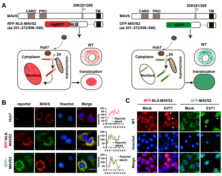
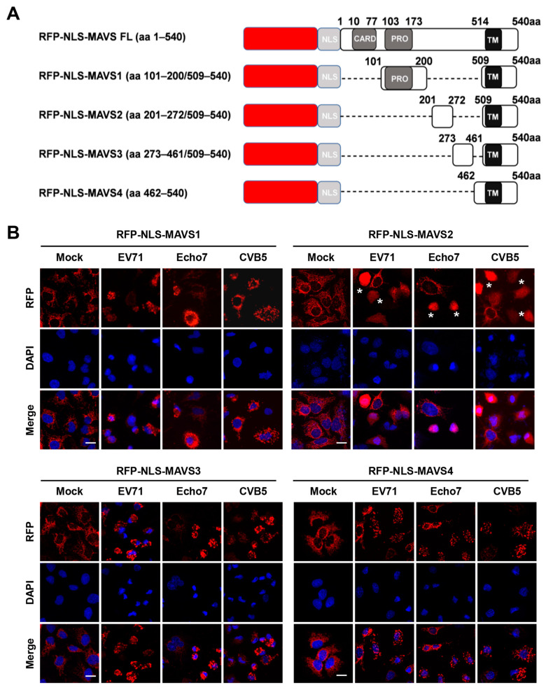
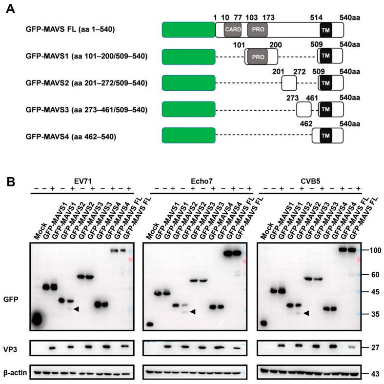
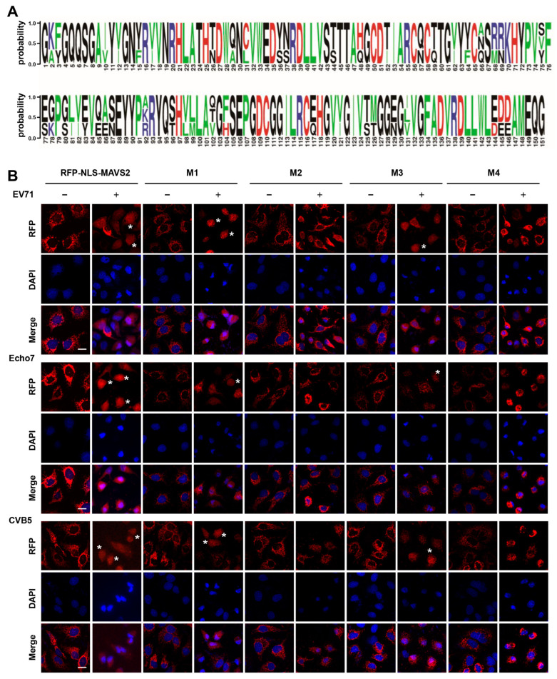
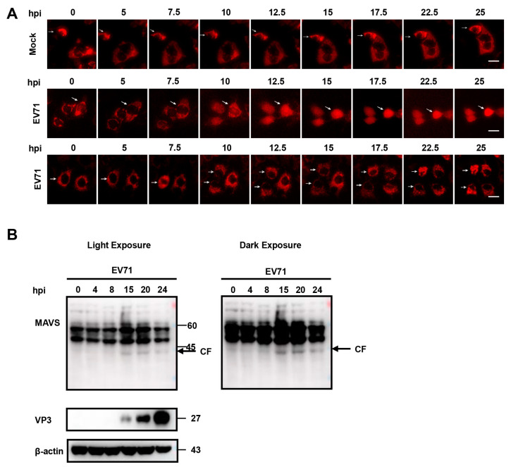
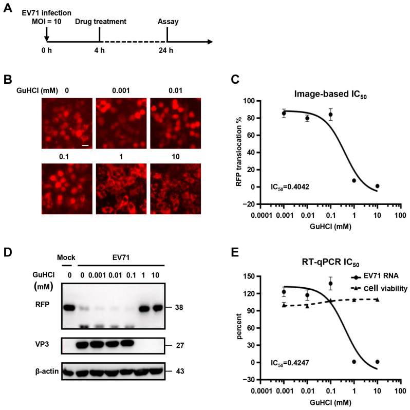
Enterovirus consists of a variety of viruses that could cause a wide range of illness in human. The pathogenesis of these viruses remains incompletely understood and no specific treatment is available. Better methods to study enterovirus infection in live cells will help us better understand the pathogenesis of these viruses and might contribute to antiviral development. Here in this study, we developed fluorescent cell-based reporter systems that allow sensitive distinction of individual cells infected with enterovirus 71 (EV71). More importantly, these systems could be easily used for live-cell imaging by monitoring viral-induced fluorescence translocation after EV71 infection. We further demonstrated that these reporter systems could be used to study other enterovirus-mediated MAVS cleavage and they are sensitive for antiviral activity testing. Therefore, integration of these reporters with modern image-based analysis has the potential to generate new insights into enterovirus infection and facilitate antiviral development.
Keywords: antiviral; enterovirus; fluorescence; live cell imaging; reporter.
Conflict of interest statement
The authors declare no conflict of interest.
Figures
Similar articles
-
In Vivo Imaging with Bioluminescent Enterovirus 71 Allows for Real-Time Visualization of Tissue Tropism and Viral Spread.J Virol. 2017 Feb 14;91(5):e01759-16. doi: 10.1128/JVI.01759-16. Print 2017 Mar 1. J Virol. 2017. PMID: 27974562 Free PMC article.
-
Antiviral effects of two Ganoderma lucidum triterpenoids against enterovirus 71 infection.Biochem Biophys Res Commun. 2014 Jul 4;449(3):307-12. doi: 10.1016/j.bbrc.2014.05.019. Epub 2014 May 15. Biochem Biophys Res Commun. 2014. PMID: 24845570
-
Development of a stable Gaussia luciferase enterovirus 71 reporter virus.J Virol Methods. 2015 Jul;219:62-66. doi: 10.1016/j.jviromet.2015.03.020. Epub 2015 Apr 2. J Virol Methods. 2015. PMID: 25843263
-
Development of antiviral agents toward enterovirus 71 infection.J Microbiol Immunol Infect. 2015 Feb;48(1):1-8. doi: 10.1016/j.jmii.2013.11.011. Epub 2014 Feb 21. J Microbiol Immunol Infect. 2015. PMID: 24560700 Review.
-
Animal models of enterovirus 71 infection: applications and limitations.J Biomed Sci. 2014 Apr 17;21(1):31. doi: 10.1186/1423-0127-21-31. J Biomed Sci. 2014. PMID: 24742252 Free PMC article. Review.
Cited by
-
Receptors and Host Factors for Enterovirus Infection: Implications for Cancer Therapy.Cancers (Basel). 2024 Sep 12;16(18):3139. doi: 10.3390/cancers16183139. Cancers (Basel). 2024. PMID: 39335111 Free PMC article. Review.
-
The class III phosphatidylinositol 3-kinase VPS34 supports EV71 replication by promoting viral replication organelle formation.J Virol. 2024 Oct 22;98(10):e0069524. doi: 10.1128/jvi.00695-24. Epub 2024 Sep 10. J Virol. 2024. PMID: 39254312 Free PMC article.
-
N-terminal acetyltransferase 6 facilitates enterovirus 71 replication by regulating PI4KB expression and replication organelle biogenesis.J Virol. 2024 Feb 20;98(2):e0174923. doi: 10.1128/jvi.01749-23. Epub 2024 Jan 8. J Virol. 2024. PMID: 38189249 Free PMC article.
-
Natural-Target-Mimicking Translocation-Based Fluorescent Sensor for Detection of SARS-CoV-2 PLpro Protease Activity and Virus Infection in Living Cells.Int J Mol Sci. 2024 Jun 17;25(12):6635. doi: 10.3390/ijms25126635. Int J Mol Sci. 2024. PMID: 38928340 Free PMC article.
References
Publication types
MeSH terms
Substances
LinkOut - more resources
Full Text Sources
Miscellaneous
