Review
. 2023 Apr 29;15(5):1088.
doi: 10.3390/v15051088.

Three-Dimensional Chromatin Structure of the EBV Genome: A Crucial Factor in Viral Infection

Affiliations
Review

Three-Dimensional Chromatin Structure of the EBV Genome: A Crucial Factor in Viral Infection

Lisa Beatrice Caruso et al. Viruses. .

Abstract

Epstein-Barr Virus (EBV) is a human gamma-herpesvirus that is widespread worldwide. To this day, about 200,000 cancer cases per year are attributed to EBV infection. EBV is capable of infecting both B cells and epithelial cells. Upon entry, viral DNA reaches the nucleus and undergoes a process of circularization and chromatinization and establishes a latent lifelong infection in host cells. There are different types of latency all characterized by different expressions of latent viral genes correlated with a different three-dimensional architecture of the viral genome. There are multiple factors involved in the regulation and maintenance of this three-dimensional organization, such as CTCF, PARP1, MYC and Nuclear Lamina, emphasizing its central role in latency maintenance.

Keywords: CTCF; EBV; chromatin looping; chromatin structure; cohesin; epigenetics; latency.


Conflict of interest statement

The authors declare no conflict of interest.

Figures
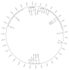
Figure 1
Figure 1
Circular view of the EBV genome. The EBV genome is represented as a circle. For better visualization, only the viral genes and promoters that will be mentioned and discussed in this review are listed.
Figure 2
Figure 2
Schematic view of the different latency types. Upon infection, EBV is able to establish latent infection in the host cell. The three main types of latency are depicted in this image. From the left: Type I latency is characterized by the expression of only the viral nuclear protein EBNA1 and the noncoding microRNAs EBERs; Type II latency involves the expression of EBNA1, EBERs, BARTs and the three transmembrane proteins LMP1, 2a, 2b. Type III latency is characterized by the expression of all the previously mentioned genes and the viral transcription factors EBNA2, EBNA3A, EBNA3B, EBNA3C, and EBNA-LP. Created with BioRender.com (accessed on 2 April 2023).
Figure 3
Figure 3
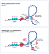
Schematic view on the 3D structure of the EBV genome. (Top) PARP1 PARylates CTCF, thus stabilizing its binding on the viral genome. CTCF, together with Cohesin, forms loops in close proximity with viral promoters with enhancers, promoting viral latent gene expression. (Bottom) PARP1 inhibition determines a reduction in CTCF binding, therefore causing a disruption of loops and a consequent decrease in viral gene expression. Created with BioRender.com (accessed on 2 April 2023).

References

    1. Epstein M.A., Achong B.G., Barr Y.M. Virus Particles in Cultured Lymphoblasts from Burkitt’s Lymphoma. Lancet. 1964;1:702–703. doi: 10.1016/S0140-6736(64)91524-7. - DOI - PubMed
    1. Young L.S., Yap L.F., Murray P.G. Epstein-Barr virus: More than 50 years old and still providing surprises. Nat. Rev. Cancer. 2016;16:789–802. doi: 10.1038/nrc.2016.92. - DOI - PubMed
    1. Dunmire S.K., Verghese P.S., Balfour H.H. Primary Epstein-Barr virus infection. J. Clin. Virol. 2018;102:84–92. doi: 10.1016/j.jcv.2018.03.001. - DOI - PubMed
    1. Gewurz B., Longnecker R., Cohen J.I. Epstein-Barr Virus. In: Howley P., Knipe D.M., Cohen J.I., Damania B., editors. Fields Virology. 7th ed. Wolters Kluwer; Philadelphia, PA, USA: 2021. pp. 324–388.
    1. Khan G., Hashim M.J. Global burden of deaths from Epstein-Barr virus attributable malignancies 1990–2010. Infect. Agents Cancer. 2014;9:38. doi: 10.1186/1750-9378-9-38. - DOI - PMC - PubMed

Publication types

LinkOut - more resources