. 2023 Apr 29;15(5):1098.
doi: 10.3390/v15051098.

Elucidation of the Cellular Interactome of African Swine Fever Virus Fusion Proteins and Identification of Potential Therapeutic Targets

Affiliations

Elucidation of the Cellular Interactome of African Swine Fever Virus Fusion Proteins and Identification of Potential Therapeutic Targets

Isabel García-Dorival et al. Viruses. .

Abstract

African swine fever virus (ASFV) encodes more than 150 proteins, most of them of unknown function. We used a high-throughput proteomic analysis to elucidate the interactome of four ASFV proteins, which potentially mediate a critical step of the infection cycle, the fusion and endosomal exit of the virions. Using affinity purification and mass spectrometry, we were able to identify potential interacting partners for those ASFV proteins P34, E199L, MGF360-15R and E248R. Representative molecular pathways for these proteins were intracellular and Golgi vesicle transport, endoplasmic reticulum organization, lipid biosynthesis, and cholesterol metabolism. Rab geranyl geranylation emerged as a significant hit, and also Rab proteins, which are crucial regulators of the endocytic pathway and interactors of both p34 and E199L. Rab proteins co-ordinate a tight regulation of the endocytic pathway that is necessary for ASFV infection. Moreover, several interactors were proteins involved in the molecular exchange at ER membrane contacts. These ASFV fusion proteins shared interacting partners, suggesting potential common functions. Membrane trafficking and lipid metabolism were important categories, as we found significant interactions with several enzymes of the lipid metabolism. These targets were confirmed using specific inhibitors with antiviral effect in cell lines and macrophages.

Keywords: ASFV; African swine fever virus; drug target; fusion proteins; interactome; lipid metabolism enzymes; virus–host interaction.


Conflict of interest statement

The authors declare that they have not known competing financial interest or personal relationship that could have appeared to influence the work reported in this paper.

Figures

Figure 9
Figure 9
Lipid traffic-related proteins and ASFV infection. Immunofluorescence confocal images of mock (upper panel) or ASFV-infected cells at 8 (intermediate panel) and 16 hpi (lower panels). Lipid traffic-related proteins SACM1L, PI4K, SREBP and TMEM33 localized at or around ASFV replication sites (VF) as stained with a monoclonal antibody against ASFV p150 protein and rabbit antibodies against: (A) TMEM33, (B) CCT4, (C) SACM1L, (D) PI4K and (E). SREBP2, respectively, at the time points indicated. Scale bar: 20 µm. Zoom: 5 µm.
Figure 10
Figure 10
ASFV infection and inhibitors of lipid metabolism in Vero cells and macrophages. Inhibition of infection efficiency in cells treated with compounds inhibiting lipid metabolism. Inhibitor or DMSO pretreated ASFV-infected Vero cells (A) or macrophages (B) for 16 hpi, as detected by flow cytometry of cells infected with B54GFP and depicted as median and standard deviations in histograms. Statistically significant data are shown with asterisks (**** p < 0.0001, *** p < 0.001 and ns non-significant). The following lipid inhibitor compounds were used: ATV, atorvastatin; EZT ezetimibe; CLZ, cilostazol, ST1326; and TC, triacsin-C.
Figure 1
Figure 1
Expression and coimmunoprecipitation of GFP-tagged ASFV proteins (GFP-VP). (A) Expression of ASFV proteins MGF360-15R, P34, E248R, E199L and control GFP in HEK 293T cells as confirmed by immunofluorescence. As expected, cells showed expression of GFP (upper panel) and diverse distribution of viral proteins GFP-MGF360-15R, GFP-P34, GFP-A278R and GFP-E199L. GFP, Topro3 and Merge images are indicated in upper panels in colors. Scale bar 25 µm. (B) WB detection of GFP-tagged ASFV viral proteins MGF360-15R, P34, E248R, E199L and control GFP in the immunoprecipitation assay. “I” refers to the input sample and “E” to the elution sample. Cell lysates from transfected cells were co-immunoprecipitated with agarose beads.
Figure 2
Figure 2
Volcano plots representing mass spectrometry and statistical analysis results for ASFV-tagged viral proteins. Analysis by MS included EGFP-tagged ASFV proteins and control. The immunoprecipitation and label–free mass spectrometry analysis was performed in triplicate. The logarithmic fold change is plotted against the negative logarithmic p values of the t test. In these volcano plots, the dashed curve indicates the region of significant interactions; the dots in the upper right quadrant represent potential protein-interacting partners for the following ASFV proteins: (A) EGFP-P34, (B) EGFP-E199L, (C) EGFP- MGF360-15R and (D) EGFP-E248R. For any potential protein interaction partner, the values of their abundance when coimmunoprecipitated with those EGFP–ASFV fusion proteins were compared to their value from the coimmunoprecipitation with the control (EGFP control). Dots corresponding to relevant hits from statistical analysis are highlighted in red.
Figure 3
Figure 3
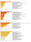
Bioinformatic and functional analysis of the ASFV interactome of proteins P34, E199L, MGF360-15R and E248R. Bar graph of enriched terms across input gene list, colored with intensities according to their p-values. Bar graph represents the top 20 enriched terms (such as GO/KEGG terms, canonical pathways, hall mark gene sets, etc.) across the different ASFV proteins, colored according to their –log10 (p-value) for proteins: (A) P34, (B) E199L, (C) MGF360-15R and (D) E248R. A complete list of the terms in each cluster can be found in the supporting information (Table S1).
Figure 4
Figure 4
Network of enriched terms colored by cluster ID of the interactome for ASFV proteins (A) P34, (B) E199L, (C) MGF360-15R and (D) E248R. To further capture the relationships between the terms, a subset of representative terms from the full cluster was selected and converted as a network layout. More specifically, each term is represented by a circle node, where its size is proportional to the number of input genes falling under that term, and its color represents its cluster identity (i.e., nodes of the same color belong to the same cluster). For clarity, labels are only shown for one term per cluster.
Figure 5
Figure 5
Visualizations of meta-analysis results based on multiple interactomes of the ASFV viral proteins (MGF360-15R, P34, E248R and E199L). (A) Circos plot shows the overlap of genes from the input gene list. On the inside, each arc represents one gene list. Dark orange color represents the genes that are shared by multiple lists and light orange color represents genes that are unique to a gene list. Purple lines link the same gene when shared by multiple gene lists. (B) Heatmap of enriched terms across the several input gene lists (MGF360-15R, P34, E248R and E199L) colored by p-values. The heatmap cells are colored by their p-values, and grey cells indicate the lack of enrichment for that term in the corresponding gene list.
Figure 6
Figure 6
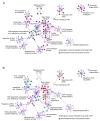
Network of enriched terms of meta-analysis results based on multiple interactomes of the ASFV viral proteins (MGF360-15R, P34, E248R and E199L). (A). Network of enriched terms represented as pie charts, where pies are color-based on the identities of the gene lists. Each pie sector is proportional to the number of hits originated from a gene list. (B). Network of enriched terms colored by cluster ID. To further capture the relationships between all the terms from the different gene lists, a subset of representative terms from the full cluster was selected and converted in a network layout.
Figure 7
Figure 7
Immunoprecipitated GFP-tagged ASFV proteins with cellular proteins. Immunoprecipitated GFP-tagged ASFV viral proteins MGF360-15R, P34, E248R, E199L (EGFP-VP) and control EGFP, with the corresponding cellular proteins in the immunoprecipitation and MS assay. (A) WB detection of GFP-tagged ASFV viral proteins EGFP-MGF360-15R, EGFP-P34, EGFP-E248R and EGFP-E199L, control EGFP and selected cellular proteins that were identified by MS analysis, in the immunoprecipitation assay. “I” refers to the input sample and “E” to the elution sample. Immunoprecipitated cellular proteins were endogenous Rab5, Rab7, Rab11, DDX3, VAPB, VAPA, Tubulin alpha and GAPDH. Proteins were found at the expected molecular weights (MW). (B) As E248R had lower expression, we loaded a larger volume of sample per well (20 µL), and we could observe that this protein immunoprecipitated cellular tubulin alpha.
Figure 8
Figure 8
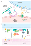
ASFV interactome significant hits related to Golgi transport and ER pathways. ER membrane contacts are important in the control of membrane trafficking and regulation of intracellular organelles. ASFV interactome significant hits are highlighted in bold letters in the schematics. (A). Upon stress and high cholesterol content, endosomes and lysosomes move to a perinuclear location clustered around the microtubule-organizing center together with vesicles of the trans-Golgi network (TGN). This movement is orchestrated by ER VAP that regulates the association (or dissociation) of ORP1L, in complex with RILP, Rab7, and ORP1L and the HOPS complex through microtubule motor dynein, as shown in panel (A). (B). Lipid transfer proteins regulate cholesterol transfer between ER and Golgi membranes, mediated by SACM1, OSBP and VAP. SACM1 is an ER-resident phosphatase that dephosphorylates PtdIns4P in the ER. It is also regulated by the carnitine palmitoyltransferase (CPT).

References

    1. Gonzales W., Moreno C., Duran U., Henao N., Bencosme M., Lora P., Reyes R., Nunez R., De Gracia A., Perez A.M. African swine fever in the Dominican Republic. Transbound. Emerg. Dis. 2021;68:3018–3019. doi: 10.1111/tbed.14341. - DOI - PubMed
    1. Tran H.T.T., Truong A.D., Dang A.K., Ly D.V., Nguyen C.T., Chu N.T., Nguyen H.T., Dang H.V. Genetic characterization of African swine fever viruses circulating in North Central region of Vietnam. Transbound. Emerg. Dis. 2021;68:1697–1699. doi: 10.1111/tbed.13835. - DOI - PubMed
    1. Dixon L.K., Chapman D.A., Netherton C.L., Upton C. African swine fever virus replication and genomics. Virus Res. 2013;173:3–14. doi: 10.1016/j.virusres.2012.10.020. - DOI - PubMed
    1. Alonso C., Miskin J., Hernaez B., Fernandez-Zapatero P., Soto L., Canto C., Rodriguez-Crespo I., Dixon L., Escribano J.M. African swine fever virus protein p54 interacts with the microtubular motor complex through direct binding to light-chain dynein. J. Virol. 2001;75:9819–9827. doi: 10.1128/JVI.75.20.9819-9827.2001. - DOI - PMC - PubMed
    1. Cuesta-Geijo M.A., Garcia-Dorival I., Del Puerto A., Urquiza J., Galindo I., Barrado-Gil L., Lasala F., Cayuela A., Sorzano C.O.S., Gil C., et al. New insights into the role of endosomal proteins for African swine fever virus infection. PLoS Pathog. 2022;18:e1009784. doi: 10.1371/journal.ppat.1009784. - DOI - PMC - PubMed

Publication types

Substances

LinkOut - more resources