. 2023 Jan-Dec;15(1):2215363.
doi: 10.1080/19420862.2023.2215363.

Molecular recognition requires dimerization of a VHH antibody

Affiliations

Molecular recognition requires dimerization of a VHH antibody

Christopher A Smith et al. MAbs. 2023 Jan-Dec.

Abstract

Camelid heavy-chain-only antibodies are a unique class of antibody that possesses only a single variable domain (termed VHH) for antigen recognition. Despite their apparent canonical mechanism of target recognition, where a single VHH domain binds a single target, an anti-caffeine VHH has been observed to possess 2:1 stoichiometry. Here, the structure of the anti-caffeine VHH/caffeine complex enabled the generation and biophysical analysis of variants that were used to better understand the role of VHH homodimerization in caffeine recognition. VHH interface mutants and caffeine analogs, which were examined to probe the mechanism of caffeine binding, suggested caffeine recognition is only possible with the VHH dimer species. Correspondingly, in the absence of caffeine, the anti-caffeine VHH was found to form a dimer with a dimerization constant comparable to that observed with VH:VL domains in conventional antibody systems, which was most stable near physiological temperature. While the VHH:VHH dimer structure (at 1.13 Å resolution) is reminiscent of conventional VH:VL heterodimers, the homodimeric VHH possesses a smaller angle of domain interaction, as well as a larger amount of apolar surface area burial. To test the general hypothesis that the short complementarity-determining region-3 (CDR3) may help drive VHH:VHH homodimerization, an anti-picloram VHH domain containing a short CDR3 was generated and characterized, which revealed it also existed as dimer species in solution. These results suggest homodimer-driven recognition may represent a more common method of VHH ligand recognition, opening opportunities for novel VHH homodimer affinity reagents and helping to guide their use in chemically induced dimerization applications.

Keywords: Camelid antibody; ITC; VHH; chemically induced dimerization (CID) systems; crystal structure; dimerization; hapten recognition; nanobody; sdAb.


Conflict of interest statement

No potential conflict of interest was reported by the authors.

Figures

Amino acid sequence alignment of VHH domains relevant to this study.
Figure 1.
Amino acid sequences of VHH domains. VHH sequences include the anti-RNase A VHH “framework” (cAbrn05) domain, the original anti-caffeine VHH by Ladenson et al. (Caff), the anti-caffeine VHH produced through grafting CDR regions (Caffgraft) examined in this study, an anti-picloram VHH (Picloram), an anti-GFP VHH (GFP), and an anti-HIV1 capsid protein C-terminal domain VHH (PDB ID: 2xV6). Grey highlighting: anti-caffeine residues introduced into segments of CDR loops of anti-RNase A VHH; underlined residues: anti-RNase A residues that remained post-grafting; bold italicized residues: residues mutated in this study to explore disruption of VHH homodimer.
Images illustrating the caffeine-binding pocket is located at the homodimer interface formed from the two VHH domains.
Figure 2.
Structure of caffeine/anti-caffeine VHH homodimer complex. (a) Homodimer stick structure highlighting caffine binding site and conserved water molecules within VHH:VHH interface. (b) Close up of caffeine binding site. Image is rotated 90° vertically from panel A. Y32 sidechains from both VHH domains, which are are shown in stick (foreground) and space fill (background), “sandwich” the caffine ligand. (c) Electron density map (2FoFc) of caffeine/CDR3-binding pocket with water mediated hydrogen bond. Map coutoured at 2σ. (d) Side and top view perspective of the VHH complex. VHH domains (white and gray); caffeine (magenta); interface mutations F47R (cyan), V100R (green), and Y(100B)R (orange); Kabat numbering is use for residue positions. Caffeine shown in magenta in all panels.
Analytical ultracentrifugation data suggesting the VHH domain exists as a monomer-dimer species in the absence of caffeine.
Figure 3.
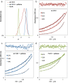
Analytical ultracentrifugation analysis of anti-caffeine VHH dimer assembly. (a) Sedimentation coefficient distribution plots for wt. VHH alone, wt VHH with stoichiometric caffeine, and F47R VHH. Each protein was loaded at a concentration of 30 µM, and the caffeine concentration for the second sample was 15 µM. (b) Sedimentation equilibrium data for wt VHH. (c) Sedimentation equilibrium data for wt VHH with stoichiometric caffeine. (d) Sedimentation equilibrium data for F47R VHH with stoichiometric caffeine. All equilibrium experiments were conducted using protein samples at 3, 10, and 30 µM, with stoichiometric levels of caffeine (1.5, 5, and 15 µM) as required. The fits shown for each sample are the result of global analysis of all three speeds and concentrations; for clarity, only the 10 µM data at each speed are plotted.
Cartoon model of linked dimerization/binding and data plots showing the dependence of caffeine’s binding enthalpy as a function of temperature and the calculated stability of the dimer as a function of temperature. The dimer species is most stable near physiological temperature.
Figure 4.
Thermodynamic model and temperature profile for anti-caffeine VHH dimer dissociation. (a) Linked thermodynamic model consisting of dimerization, Kdimer, followed by caffeine binding to the VHH dimer species, Kbind. (b) Plot of ΔH°obs as a function of temperature for VHH:VHH dissociation. (c) Plot of ΔG°obs as a function of temperature for VHH:VHH dissociation based on obtained dimer dissociation thermodynamic parameters. Includes data from AUC experiments at 20° for comparison. Error bars represent 95% confidence intervals. Note: Thermodynamic terms for ΔH°obs and ΔG°obs presented in panels B and C represent the dimer dissociation reaction, which is the opposite direction of Kdimer presented in panel A.
Image highlighting that the VHH homodimer buries more surface area and is more parallel in VHH:VHH domain alignment, as compared to conventional VH/VL heterodimers.
Figure 5.
Structural differences between the anti-caffeine VHH homodimer and conventional VH/VL heterodimers. (a) Surface area burial heat map for residues within the anti-caffeine VHH:VHH (left) and a representative VH:VL dimer (right; PDB ID: 1Q72). (b) Structural overlay of a VHH domain from the anti-caffeine structure with the VH domain from a murine anti-cocaine Fab (PDB entry 1Q72). The VH and VHH domain alignment is in background (gray) and the VL domain from anti-cocaine (blue) and second VHH domain from anti-caffeine VHH (red) are in foreground. Average angle between principal axes is calculated from four anti-caffeine dimers and a sampling of VH:VL structures from the PDB 1Q72, 2JB6, 2UUD, 3CFB, 3FO9).
Image indicating the short CDR3 of the anti-caffeine VHH would be expected to expose significantly more framework surface area, as compared to a conventional VHH domain.
Figure 6.
Stereoview image displaying the difference in CDR3 conformation between anti-caffeine VHH domain and a conventional anti-RNase a VHH. VHH domains have been superimposed and anti-RNase a framework omitted for clarity. CDR3 loop color coded: anti-caffeine VHH (red) and anti-RNaseA VHH (cyan). Framework residues that experience greater solvent exposure due to displaced CDR3 are highlighted in green sticks. Caffeine ligand is displayed in stick form at top of image (carbon-green).
Size exclusion chromatography plot which illustrate anti-picloram VHH appears as a homodimer in solution in the absence of picloram ligand.
Figure 7.
Normalized size exclusion profile of anti-picloram versus monomeric anti-RNase a VHH domains. Anti-RNase a (dashed pink) and anti-picloram VHH (solid blue).

References

    1. Hamers-Casterman C, Atarhouch T, Muyldermans S, Robinson G, Hamers C, Songa EB, Bendahman N, Hamers R.. Naturally occurring antibodies devoid of light chains. Nature. 1993;363(6428):446–14. PMID: 8502296. doi:10.1038/363446a0. - DOI - PubMed
    1. Spinelli S, Tegoni M, Frenken L, van Vliet C, Cambillau C. Lateral recognition of a dye hapten by a llama VHH domain. J Mol Biol. 2001;311(1):123–29. PMID: 11469862. doi:10.1006/jmbi.2001.4856. - DOI - PubMed
    1. Muyldermans S, Atarhouch T, Saldanha J, Barbosa JA, Hamers R. Sequence and structure of VH domain from naturally occurring camel heavy chain immunoglobulins lacking light chains. Protein Eng. 1994;7(9):1129–35. PMID: 7831284. doi:10.1093/protein/7.9.1129. - DOI - PubMed
    1. Wu TT, Johnson G, Kabat EA. Length distribution of CDRH3 in antibodies. Proteins: Struct Funct Genet. 1993;16(1):1–7. PMID: 8497480. doi:10.1002/prot.340160102. - DOI - PubMed
    1. Decanniere K, Desmyter A, Lauwereys M, Ghahroudi MA, Muyldermans S, Wyns L. A single-domain antibody fragment in complex with RNase A: non-canonical loop structures and nanomolar affinity using two CDR loops. Structure. 1999;7(4):361–70. PMID: 10196124. doi:10.1016/s0969-2126(99)80049-5. - DOI - PubMed

Publication types

MeSH terms

Substances

LinkOut - more resources