

Novel discovery of a lymphatic bridge connecting Schlemm's canal to limbal and conjunctival lymphatic pathway
- PMID: 37244593
- PMCID: PMC10567112
- DOI: 10.1016/j.jtos.2023.05.009
Novel discovery of a lymphatic bridge connecting Schlemm's canal to limbal and conjunctival lymphatic pathway
Abstract
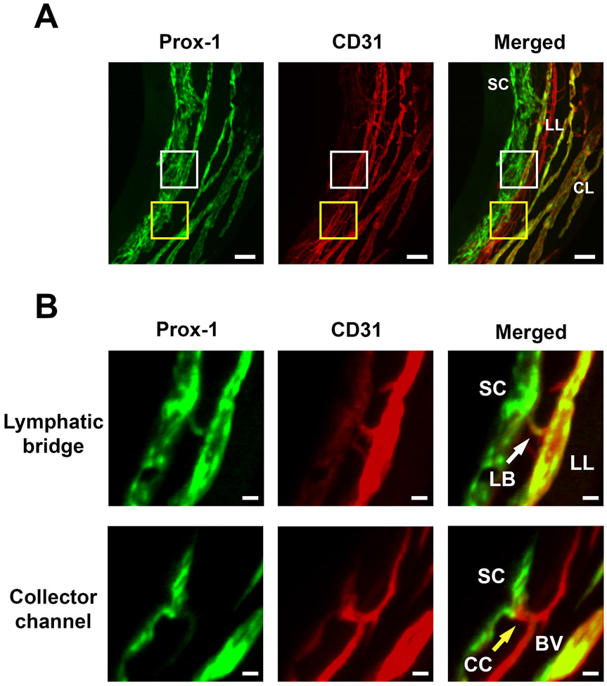
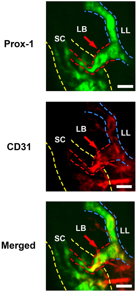
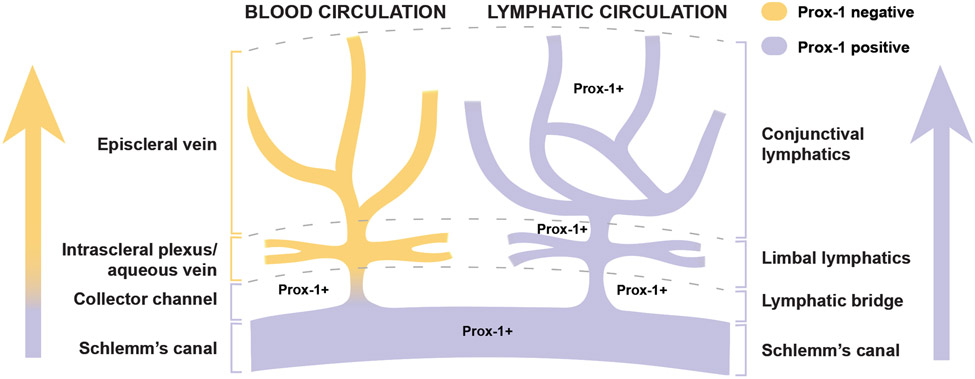
Purpose: Schlemm's canal (SC) is a critical structure regulating aqueous humor (AH) drainage and intraocular pressure (IOP). It is known that in the conventional outflow pathway, AH flows from SC to episcleral veins. We recently reported a high-resolution three-dimensional (3D) imaging technology for intact eyeballs, SC and ocular surface. Using this advanced technology, we herein report the discovery of a new structure, termed lymphatic bridge, that directly connects SC to the limbal and conjunctival lymphatic pathway. Further investigation on this novel outflow pathway may provide new mechanisms and therapeutic approaches for glaucoma.
Methods: As reported previously, intact eyeballs were harvested from Prox-1-GFP (green fluorescent protein) mice and processed by a tissue clearing technique with CLARITY. Samples were immunolabeled with specific antibodies for CD31 (pan-endothelial marker) and LYVE-1 (lymphatic vessel endothelial hyaluronan receptor-1) and imaged by light-sheet fluorescent microscopy. The limbal areas were examined to locate connecting channels between SC and limbal and conjunctival lymphatic vessels. Moreover, in vivo anterior chamber dye injection was performed with Texas Red dextran for functional analysis on AH outflow.
Results: A novel lymphatic bridge structure that expressed both Prox-1 and LYVE-1 was discovered between the SC and limbal lymphatic vessels connected with conjunctival lymphatic pathway. Results from the anterior chamber dye injection also confirmed AH drainage into the conjunctival lymphatic outflow pathway.
Conclusions: This study provides the first evidence on the direct connection between SC and conjunctival lymphatic pathway. This new pathway is distinctive from the traditional episcleral vein pathway and merits further investigation.
Keywords: Aqueous humor drainage pathway; Conjunctiva; Limbus; Lymphatic vessel; Schlemm's canal.
Copyright © 2023 Elsevier Inc. All rights reserved.
Conflict of interest statement
Declaration of competing interest None.
Figures


Similar articles
-
Schlemm's canal: the outflow 'vessel'.Acta Ophthalmol. 2022 Jun;100(4):e881-e890. doi: 10.1111/aos.15027. Epub 2021 Sep 13. Acta Ophthalmol. 2022. PMID: 34519170 Free PMC article. Review.
-
Novel characterization and live imaging of Schlemm's canal expressing Prox-1.PLoS One. 2014 May 14;9(5):e98245. doi: 10.1371/journal.pone.0098245. eCollection 2014. PLoS One. 2014. PMID: 24827370 Free PMC article.
-
The three-dimensional organisation of the post-trabecular aqueous outflow pathway and limbal vasculature in the mouse.Exp Eye Res. 2014 Aug;125:226-35. doi: 10.1016/j.exer.2014.06.011. Epub 2014 Jun 27. Exp Eye Res. 2014. PMID: 24979218
-
Aqueous humor as eye lymph: A crossroad between venous and lymphatic system.Exp Eye Res. 2024 Jun;243:109904. doi: 10.1016/j.exer.2024.109904. Epub 2024 Apr 18. Exp Eye Res. 2024. PMID: 38642600 Review.
-
Transcriptomic profiling of Schlemm's canal cells reveals a lymphatic-biased identity and three major cell states.Elife. 2024 Oct 18;13:RP96459. doi: 10.7554/eLife.96459. Elife. 2024. PMID: 39422453 Free PMC article.
Cited by
-
In vivo quantification of human aqueous veins by enhanced depth imaging optical coherence tomography and optical coherence tomography angiography images.Int J Ophthalmol. 2023 Sep 18;16(9):1482-1488. doi: 10.18240/ijo.2023.09.15. eCollection 2023. Int J Ophthalmol. 2023. PMID: 37724266 Free PMC article.
-
Prox1 Protein in Corneal Limbal Lymphatic Vessels Maintains Limbal Stem Cell Stemness and Regulates Corneal Injury Repair.Invest Ophthalmol Vis Sci. 2025 Apr 1;66(4):81. doi: 10.1167/iovs.66.4.81. Invest Ophthalmol Vis Sci. 2025. PMID: 40298888 Free PMC article.
-
Ontogenesis of the Mouse Ocular Surface Lymphatic Vascular Network.Invest Ophthalmol Vis Sci. 2023 Dec 1;64(15):7. doi: 10.1167/iovs.64.15.7. Invest Ophthalmol Vis Sci. 2023. PMID: 38054922 Free PMC article.
-
Lymphatic vessel: origin, heterogeneity, biological functions, and therapeutic targets.Signal Transduct Target Ther. 2024 Jan 3;9(1):9. doi: 10.1038/s41392-023-01723-x. Signal Transduct Target Ther. 2024. PMID: 38172098 Free PMC article. Review.
References
-
- Cook C, Foster P. Epidemiology of glaucoma: what's new? Can J Ophthalmol. 2012;47:223–6. - PubMed
-
- Tham YC, Li X, Wong TY, Quigley HA, Aung T, Cheng CY. Global prevalence of glaucoma and projections of glaucoma burden through 2040: a systematic review and meta-analysis. Ophthalmology. 2014;121:2081–90. - PubMed
-
- Maepea O, Bill A. Pressures in the juxtacanalicular tissue and Schlemm's canal in monkeys. Exp Eye Res. 1992;54:879–83. - PubMed
Publication types
MeSH terms
Grants and funding
LinkOut - more resources
Full Text Sources
Molecular Biology Databases
Research Materials
Miscellaneous
