Review
. 2023 May 27;27(1):206.
doi: 10.1186/s13054-023-04496-5.

Hypovolemia with peripheral edema: What is wrong?

Affiliations
Review

Hypovolemia with peripheral edema: What is wrong?

Randal O Dull et al. Crit Care. .

Abstract

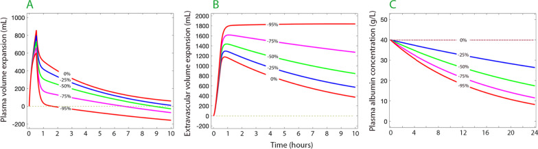
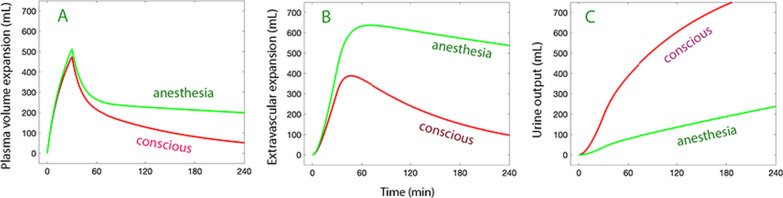
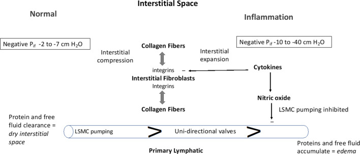
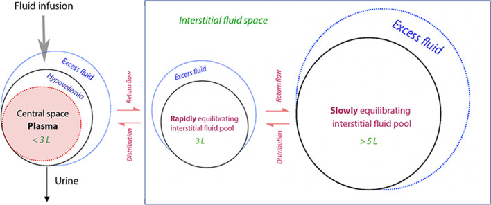
Fluid normally exchanges freely between the plasma and interstitial space and is returned primarily via the lymphatic system. This balance can be disturbed by diseases and medications. In inflammatory disease states, such as sepsis, the return flow of fluid from the interstitial space to the plasma seems to be very slow, which promotes the well-known triad of hypovolemia, hypoalbuminemia, and peripheral edema. Similarly, general anesthesia, for example, even without mechanical ventilation, increases accumulation of infused crystalloid fluid in a slowly equilibrating fraction of the extravascular compartment. Herein, we have combined data from fluid kinetic trials with previously unconnected mechanisms of inflammation, interstitial fluid physiology and lymphatic pathology to synthesize a novel explanation for common and clinically relevant examples of circulatory dysregulation. Experimental studies suggest that two key mechanisms contribute to the combination of hypovolemia, hypoalbuminemia and edema; (1) acute lowering of the interstitial pressure by inflammatory mediators such as TNFα, IL-1β, and IL-6 and, (2) nitric oxide-induced inhibition of intrinsic lymphatic pumping.


Conflict of interest statement

The authors declare that they have no competing interests.

Figures

Fig. 1
Fig. 1
Computer simulated volume expansion of the plasma (A), extravascular space (B) and the plasma albumin concentration (C) in response to infusion of 30 mL/kg over 30 min followed by 15 mL/kg over 1 h of Ringer´s acetate/lactate. The percentages imply the degree of inflicted restriction of lymphatic return of fluid and albumin. The albumin kinetics assumes a capillary leakage of 5% of the intravascular pool per hour which, at steady state, is fully returned by lymphatic flow, and is unaffected by fluid loading. The volume kinetic parameters were taken from a mixed population of conscious and anesthetized patients [4]. Each gram of albumin was modeled to bind 10 mL of fluid [5]. The extravascular volume expansion could not be divided into a fast and slowly exchanging pool because it is unknown from which pool the lymph originates
Fig. 2
Fig. 2
A Human dermis histology demonstrating hyaluronan staining (HA, brown), vascular endothelial cell of capillaries, small arteries and veins (CD34, magenta) and lymphatic vessels (LYVE-1, teal). Note intercellular HA in epidermis and abundant HA filling interstitial space in papillary dermis. Lymphatic vessels (*) are common in papillary dermis. B Reticular dermis. Dense HA staining is seen around peri-vascular bundles and filling inter-cellular space. Lymphatics (*) are much less common in reticular dermis. E = epidermis, PD = papillary dermis, RD = reticular dermis. Courtesy by Dr. Neil Theise and coworkers, experiments are explained in references 13 and 14
Fig. 3
Fig. 3
Distribution of an intravenous infusion of 15 ml/kg of buffered Ringer´s solution over 30 min followed by 10 mL/kg over 60 min between (A) the plasma (B) extravascular space, and (C) urine in a human weighing 65 kg (total volume 1620 mL) depending on whether he/she is conscious or under general anesthesia. Simulation based on volume kinetic parameters from 157 conscious volunteers and 85 anesthetized patients [10]. The extravascular expansion is the sum of the rapidly and the slowly equilibrating pool of fluid
Fig. 4
Fig. 4
The interstitial space is represented in its Normal state (left side) and during Inflammation (right side). The interstitial space is normally contracted by integrin-dependent binding between interstitial fibroblasts and collagen fibers (tension indicated by double-headed arrows) that induces Interstitial Compression. In the Normal state, the interstitial space is kept “dry” by the action of lymphatic clearance and baseline pressure (Pif) is slightly negative in the range of −2 to −7 cm H2O. Lymphatic clearance is dependent on activity of lymphatic smooth muscle cell pumping and functional uni-directional valves. During Inflammation, cytokines induce fibroblast-dependent relaxation and disassembly of the integrin-collagen complex, that promotes Interstitial Expansion and a more negative pressure (-10 to −40 cm H2O). Nitric oxide produced during inflammation has direct inhibitory actions on lymphatic smooth muscle cells to reduce the rate and force of lymphatic smooth muscle contraction; the reduced lymphatic clearance of fluid and proteins results in edema (bottom right)
Fig. 5
Fig. 5
The volume kinetic model used the distinguish between two interstitial fluid pools. Hypovolemia might develop if distribution to the infused fluid to the interstitial pools is prominent

References

    1. Guyton AC, Hall JE. The body fluid compartments: extracellular and intracellular fluids; interstitial fluid and edema. In: Textbook of medical physiology, 9th edn. Philadelphia: W.B. Saunders Company; 1996. p. 311–312.
    1. Hahn RG, Dull RO. Interstitial washdown and vascular albumin refill during fluid infusion: novel kinetic analysis from three clinical trials. Intensive Care Med Exp. 2021;9:44. doi: 10.1186/s40635-021-00407-6. - DOI - PMC - PubMed
    1. Hahn RG. Understanding volume kinetics. Acta Anaesthesiol Scand. 2020;64:570–578. doi: 10.1111/aas.13533. - DOI - PubMed
    1. Hahn RG. Arterial pressure and the rate of elimination of crystalloid fluid. Anesth Analg. 2017;124:1824–1833. doi: 10.1213/ANE.0000000000002075. - DOI - PubMed
    1. Lamke L-O, Liljedahl S-O. Plasma volume expansion after infusion of 5%, 20% and 25% albumin solutions in patients. Resuscitation. 1976;5:85–92. doi: 10.1016/0300-9572(76)90028-9. - DOI - PubMed

Substances

LinkOut - more resources