Review
. 2023 Sep;14(5):1085-1110.
doi: 10.1016/j.advnut.2023.05.013. Epub 2023 May 27.

β-Sitosterol as a Promising Anticancer Agent for Chemoprevention and Chemotherapy: Mechanisms of Action and Future Prospects

Affiliations
Review

β-Sitosterol as a Promising Anticancer Agent for Chemoprevention and Chemotherapy: Mechanisms of Action and Future Prospects

Haoyu Wang et al. Adv Nutr. 2023 Sep.

Abstract

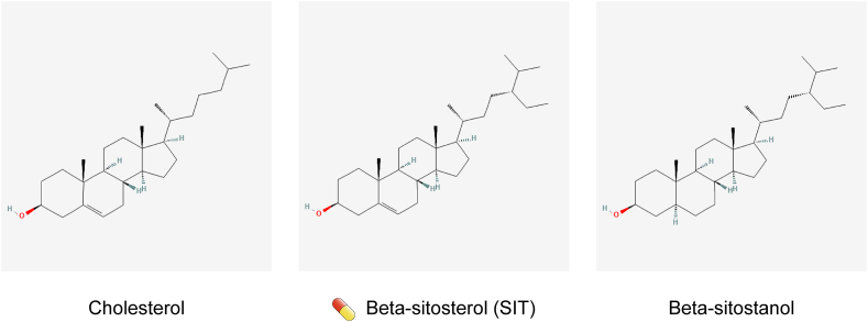
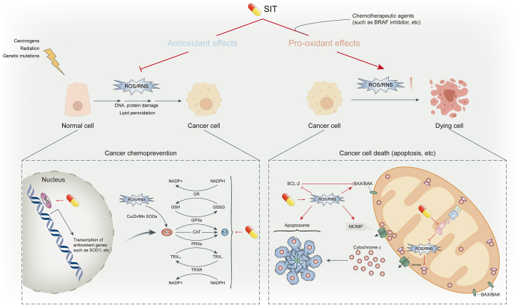
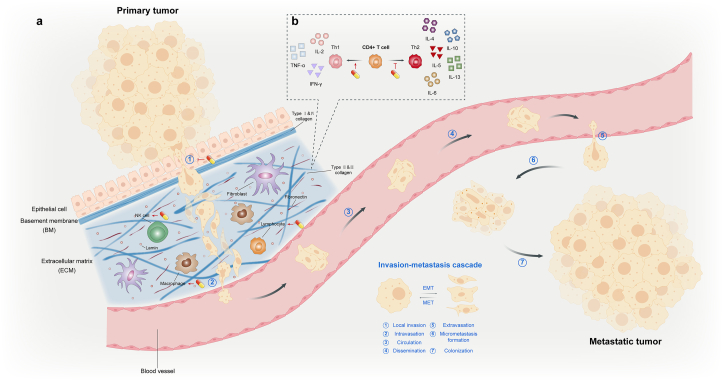
Cancer is one of the primary causes of death worldwide, and its incidence continues to increase yearly. Despite significant advances in research, the search for effective and nontoxic preventive and therapeutic agents remains greatly important. Cancer is a multimodal disease, where various mechanisms play significant roles in its occurrence and progression. This highlights the need for multitargeted approaches that are not only safe and inexpensive but also provide effective alternatives for current therapeutic regimens. β-Sitosterol (SIT), the most abundant phytosterol found in various plant foods, represents such an option. Preclinical evidence over the past few decades has overwhelmingly shown that SIT exhibits multiple anticancer activities against varied cancers, such as liver, cervical, colon, stomach, breast, lung, pancreatic, and prostate cancers, in addition to leukemia, multiple myeloma, melanoma, and fibrosarcoma. In this article, we present the latest advances and perspectives on SIT-systematically summarizing its antitumor mechanisms of action into 7 main sections and combining current challenges and prospects-for its use as a promising agent for cancer prevention and treatment. In particular, SIT plays a role in cancer prevention and treatment mainly by enhancing apoptosis, inducing cell cycle arrest, bidirectionally regulating oxidative stress, improving metabolic reprogramming, inhibiting invasion and metastasis, modulating immunity and inflammation, and combating drug resistance. Although SIT holds such great promise, the poor aqueous solubility and bioavailability coupled with low targeting efficacy limit its therapeutic efficacy and clinical application. Further research on novel drug delivery systems may improve these deficiencies. Overall, through complex and pleiotropic mechanisms, SIT has good potential for tumor chemoprevention and chemotherapy. However, no clinical trials have yet proven this potential. This review provides theoretical basis and rationality for the further design and conduct of clinical trials to confirm the anticancer activity of SIT.

Keywords: cancer; chemoprevention; chemotherapy; phytochemical; phytosterol; β-sitosterol.


Figures

Image 1
Graphical abstract
FIGURE 1
FIGURE 1
Chemical structure depictions of cholesterol, β-sitosterol (SIT), and β-sitostanol. The capsule-like symbol represents SIT.
FIGURE 2
FIGURE 2
Apoptotic signaling pathways. Apoptosis can occur through 2 pathways: extrinsic (part a) and intrinsic (part b). The extrinsic (also called death receptor) apoptotic pathway involves the binding of a death receptor ligand to a member of the death receptor family. The intrinsic (also called mitochondrial) apoptotic pathway is induced by several different stimuli that unbalance the apoptotic rheostat and lead to mitochondrial outer membrane penetration (MOMP). The extrinsic and intrinsic pathways recruit and activate the initiator caspases 8 and 9, respectively, which cleaves and activates the executioner caspases 3 and 7 to complete wide-scale cleavage of cellular components and rapid cell death. Caspase 8–mediated cleavage and activation of BH3-only protein BID (to generate tBID) connects the extrinsic pathway to the intrinsic pathway. β-Sitosterol (SIT) promotes posttranslational activation of caspase 3/7/8/9, upregulates proapoptotic members and downregulates antiapoptotic members of the BCL-2 protein family by activating p53, blocks the inhibitory effect of IAPs on caspases, increases the expression of death receptors, and acts as a sensitizer for TRAIL-induced apoptosis. Additional cell death processes, such as autophagy and anoikis, are able to modulate apoptotic signaling pathways and are affected by SIT. The capsule-like symbol represents SIT. BCL, B cell lymphoma; IAP, inhibitor of apoptosis proteins; TRAIL, TNF –related apoptosis-inducing ligand.
FIGURE 3
FIGURE 3
Cell division cycle and cell cycle control. Rings: The blue ring shows the cell division cycle, such as interphase (light blue) and M phase (dark blue). The peripheral orange-yellow ring shows the specific division of interphase and M phase and 2 decision windows. Outside the rings: Cyclin CDKs drives cell cycle progression. Under favorable conditions, the accumulation of cyclin D-CDK4/6 activity allows entry into the cell cycle, thereby preventing cell cycle exit. E2F-dependent transcription leads to the accumulation of cyclins E and A, which creates a decision window to enter S phase. Cyclin E-CDK2 activity further activates E2F-dependent transcription, forming a positive feedback loop that leads to increased activities of cyclin E-CDK2 and cyclin A-CDK2. This process allows the accumulation of cyclin A-CDK2 activity and S entry through the inactivation of APC/CCDH1 activity (not shown). Subsequent accumulation of cyclin A/B-CDK1 creates the second decision window for mitotic entry. Accumulation of cyclin A/B-CDK1 activity drives mitotic entry and allows APC/CCDC20 activation, which is required for mitotic exit and targeted degradation of cyclins to complete the cell cycle. β-Sitosterol (SIT) has an inhibitory effect on the activity of various cyclin–CDK complexes, such as cyclin D and CDK2, 4, and 6. Inside the rings: In response to DNA damage during interphase, replication stress during S phase, and abnormal spindle assembly during M phase, specific cell cycle checkpoints block or slow down the cell cycle by inhibiting CDKs activity and the APC/C. The capsule-like symbol represents SIT. APC/C, anaphase-promoting complex/cyclosome.
FIGURE 4
FIGURE 4
Antioxidant and pro-oxidant effects of β-sitosterol (SIT). The cancer preventive role of SIT is seen in its antioxidant potential, which is able to suppress abnormal ROS/RNS levels in normal cells induced by carcinogens, radiation, genetic mutations, and so on. By scavenging free radicals (not shown) and activating cellular antioxidant defense mechanisms, SIT mitigates oxidative stress and inhibits ROS/RNS-induced macromolecular damage and lipid peroxidation, thereby preventing malignant transformation of normal cells. On the contrary, in the context of cancer treatment, SIT, alone or in combination with chemotherapeutic agents, promotes the excessive accumulation of ROS/RNS by inhibiting ROS/RNS clearance systems and the mitochondrial respiratory chain. SIT-induced excessive oxidative stress causes DNA damage and activates apoptosis-related signals, prompting cancer cell death. The capsule-like symbol represents SIT. ROS, reactive oxygen species; RNS, reactive nitrogen species.
FIGURE 5
FIGURE 5
Regulation of metabolic reprogramming and cell membrane structure. Left panel (cholesterol metabolism): β-sitosterol (SIT) inhibits de novo cholesterol biosynthesis (①) by inhibiting HMGCR or other enzymes in the mevalonate pathway. SIT inhibits LDLR-mediated cholesterol uptake by directly downregulating membrane LDLR expression (②) or by activating LXRs to promote IDOL transcription (⑦). SIT decreases the expression of NPCL1 transporter (③) which is responsible for cholesterol transport. As an agonist of LXRs, SIT promotes the expression of LXR target genes ABC transporters (⑦), thereby increasing cholesterol efflux (④). In addition, SIT has a weak estrogenic effect (⑤). SIT has no influence on the cholesterol esterification process (⑥). Middle panel (sphingolipid metabolism): SIT promotes de novo CER synthesis by increasing the activity of SPT (①). The influence of SIT on CER catabolism and the salvage synthesis pathway is unclear (②). SIT induces an increase in CER production and activates PP2A, which promotes apoptosis by regulating molecules in the apoptosis pathway (③). In particular, SIT alters intracellular levels of sphingolipids and cholesterol, which are essential components of membrane lipid rafts. Therefore, receptors and signaling pathways associated with membrane lipid rafts may be significantly affected by SIT. Right panel (glucose metabolism): Although when oxygen is plentiful, tumor cells rely heavily on glycolysis to produce energy, a phenomenon known as the “Warburg effect.” SIT has an antiglycolytic effect on cancer cells. The capsule-like symbol represents SIT. ABC, ATP-binding cassette; CER, ceremide; HMGCR, 3-hydroxy-3-methylglutaryl-CoA reductase; IDOL, inducible degrader of the LDLR; LDLR, LDL receptor; LXR, liver X receptor; NPCL1, Niemann-Pick C1-like 1; PP2A, protein phosphatase 2A; SPT, serine palmitoyltransferase.
FIGURE 6
FIGURE 6
Invasion–metastasis cascade and tumor microenvironment. The invasion–metastasis cascade is a series of biological events that occur during tumor metastasis, such as the departure from primary growth sites, translocation in the systemic circulation, and survival and colonization in distant tissues. The invasion–metastasis cascade can be subdivided into 7 processes. Meanwhile, EMT and its counter-process MET play an important role in tumor metastasis. On the one hand, β-sitosterol (SIT) prevents tumor cells from acquiring a mesenchymal phenotype and detaching from surrounding cells by inhibiting EMT. To the contrary, SIT inhibits the erosion of the BM and ECM by tumor cells. Taken together, SIT mainly blocks the invasion–metastasis cascade in the initial process (part a). Whether during metastasis or at primary/distant colonization sites, immune cells are an important component of the TME and play a tumor-suppressing or tumor-promoting role. SIT promotes lymphocyte proliferation and strengthens the activity of a variety of immune cells, such as NK cells and macrophages. The regulation of immune cells by SIT enhances the effect of killing tumors and inhibiting metastasis. In addition, SIT is able to shift the TH1/TH2 balance toward TH1, which is abnormally inclined toward TH2 in the TME. Thus, antitumor effects of TH1 cytokines are enhanced and tumor-promoting effects of TH2 cytokines weakened (part b). The capsule-like symbol represents SIT. BM, basement membrane; ECM, extracellular matrix; EMT, epithelial-mesenchymal transition; MET, mesenchymal–epithelial transition; TH, T-helper subset; TME, tumor microenvironment.
FIGURE 7
FIGURE 7
Active isoprenoid biosynthesis–induced MDR phenotype. Cancer cells, particularly drug-resistant cells, exhibit a high flux through the mevalonate pathway. The mevalonate pathway is the main pathway for cholesterol biosynthesis, and isoprenoid moieties, such as FPP and GGPP, are byproducts of this pathway. Farnesylation and geranylgeranylation determine the activation of Ras and RhoA proteins that engage their downstream transducers, ERK1/2 and RhoA kinase, respectively, to phosphorylate and activate transcription factor HIF. HIF-1α upregulates MDR1 and glycolytic genes, increasing the amount of P-gp and the amount of intracellular ATP produced by glycolysis. Consequently, the activity of several drug efflux transporters—P-gp, BCRP, and MRPs—is increased. This complex mechanism network is druggable by β-sitosterol (SIT), and these MDR transporters themselves can be directly inhibited by SIT as well. BCRP, breast cancer resistance protein; ERK, extracellular regulatory kinase; FPP, farnesyl pyrophosphate; GGPP, geranylgeranyl pyrophosphate; HIF, hypoxia-induced factor; MDR, multidrug resistance; MRP, multidrug resistance–associated protein; P-gp, P-glycoprotein.

References

    1. Wu C., Li M., Meng H., Liu Y., Niu W., Zhou Y., et al. Analysis of status and countermeasures of cancer incidence and mortality in China. Sci. China Life. Sci. 2019;62:640–647. - PubMed
    1. Sung H., Ferlay J., Siegel R.L., Laversanne M., Soerjomataram I., Jemal A., et al. Global cancer statistics 2020: GLOBOCAN estimates of incidence and mortality worldwide for 36 cancers in 185 countries. C.A. Cancer J. Clin. 2021;71:209–249. - PubMed
    1. Buyel J.F. Plants as sources of natural and recombinant anti-cancer agents. Biotechnol. Adv. 2018;36:506–520. - PubMed
    1. Nasir A., Bullo M.M.H., Ahmed Z., Imtiaz A., Yaqoob E., Jadoon M., et al. Nutrigenomics: epigenetics and cancer prevention: a comprehensive review. Crit. Rev. Food Sci. Nutr. 2020;60:1375–1387. - PubMed
    1. Moreau R.A., Whitaker B.D., Hicks K.B. Phytosterols, phytostanols, and their conjugates in foods: structural diversity, quantitative analysis, and health-promoting uses. Prog. Lipid Res. 2002;41:457–500. - PubMed