. 2023 Jul;29(7):1775-1781.
doi: 10.1038/s41591-023-02380-x. Epub 2023 May 29.

Astrocyte reactivity influences amyloid-β effects on tau pathology in preclinical Alzheimer's disease

Affiliations

Astrocyte reactivity influences amyloid-β effects on tau pathology in preclinical Alzheimer's disease

Bruna Bellaver et al. Nat Med. 2023 Jul.

Abstract

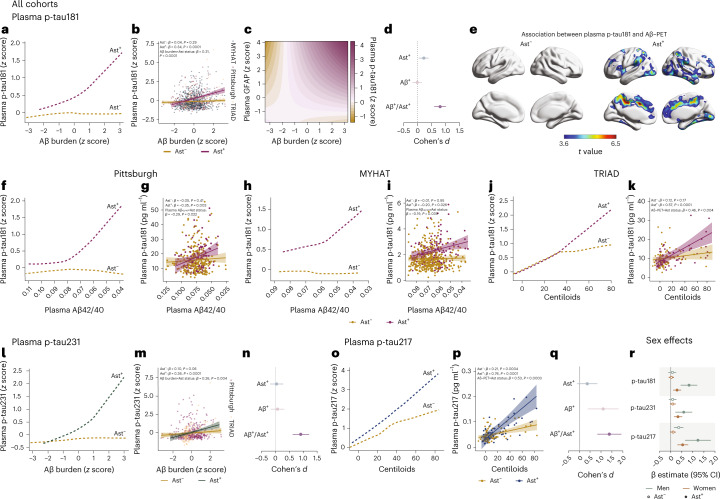
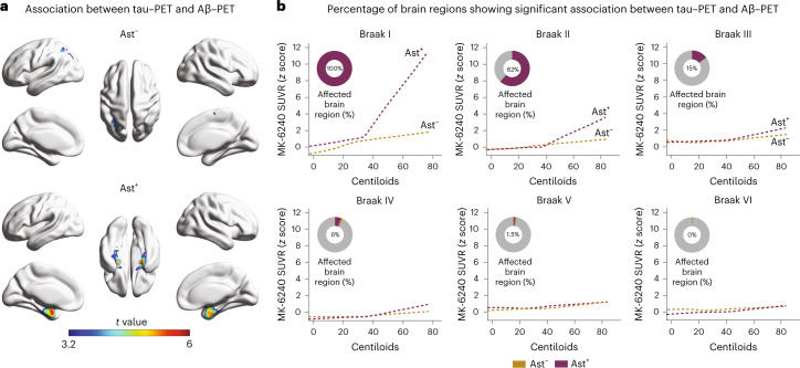
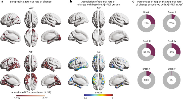
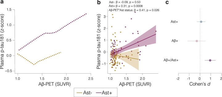
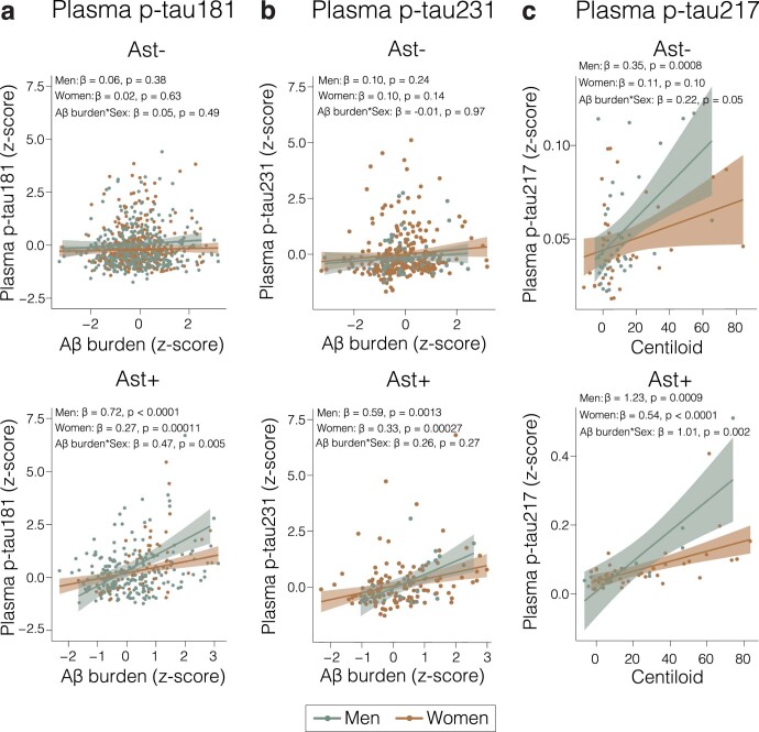
An unresolved question for the understanding of Alzheimer's disease (AD) pathophysiology is why a significant percentage of amyloid-β (Aβ)-positive cognitively unimpaired (CU) individuals do not develop detectable downstream tau pathology and, consequently, clinical deterioration. In vitro evidence suggests that reactive astrocytes unleash Aβ effects in pathological tau phosphorylation. Here, in a biomarker study across three cohorts (n = 1,016), we tested whether astrocyte reactivity modulates the association of Aβ with tau phosphorylation in CU individuals. We found that Aβ was associated with increased plasma phosphorylated tau only in individuals positive for astrocyte reactivity (Ast+). Cross-sectional and longitudinal tau-positron emission tomography analyses revealed an AD-like pattern of tau tangle accumulation as a function of Aβ only in CU Ast+ individuals. Our findings suggest astrocyte reactivity as an important upstream event linking Aβ with initial tau pathology, which may have implications for the biological definition of preclinical AD and for selecting CU individuals for clinical trials.


Conflict of interest statement

H.C.K. and G.T.-B. are employees and stockholders of Johnson & Johnson. H.Z. has served on scientific advisory boards and/or as a consultant for Abbvie, Acumen, Alector, Alzinova, ALZPath, Annexon, Apellis, Artery Therapeutics, AZTherapies, CogRx, Denali, Eisai, Nervgen, Novo Nordisk, Optoceutics, Passage Bio, Pinteon Therapeutics, Prothena, Red Abbey Labs, reMYND, Roche, Samumed, Siemens Healthineers, Triplet Therapeutics and Wave; has given lectures in symposia sponsored by Cellectricon, Fujirebio, Alzecure, Biogen and Roche and is a cofounder of Brain Biomarker Solutions in Gothenburg AB (BBS), which is a part of the GU Ventures Incubator Program (outside submitted work). K.B. has served as a consultant, at advisory boards, or at data monitoring committees for Abcam, Axon, Biogen, JOMDD/Shimadzu, Julius Clinical, Lilly, MagQu, Novartis, Prothena, Roche Diagnostics and Siemens Healthineers and is a cofounder of Brain Biomarker Solutions in Gothenburg AB (BBS), which is a part of the GU Ventures Incubator Program, all unrelated to the work presented in this paper. S.G. has served as a scientific advisor to Cerveau Therapeutics. E.R.Z. serves on the scientific advisory board of Next Innovative Therapeutics (Nintx). P.R.-N. has served on scientific advisory boards and/or as a consultant for Eisai, Novo Nordisk and Roche. N.J.A. has given lectures in symposia sponsored by Lilly and Quanterix. M.D.I. has received research funding from GE Healthcare and Avid Radiopharmaceuticals. R.C.T. serves as a consultant, advisor and/or on the medical advisory board of Happify Health, Astellas Pharma, Bayer and Hello Therapeutics. GE Healthcare holds a license agreement with the University of Pittsburgh based on the PiB PET technology described in this paper. W.E.K. is a co-inventor of PiB and, as such, has a financial interest in this license agreement. GE Healthcare provided no grant support for this study and had no role in the design or interpretation of results or preparation of this paper. The other authors declare no competing interests.

Figures

Fig. 1
Fig. 1. Astrocyte reactivity influences Aβ-dependent tau phosphorylation.
a, Robust linear regressions show that plasma p-tau181 increases as a function of Aβ burden only in the presence of astrocyte reactivity (Ast+) in all cohorts together (n = 1,016). b, Linear regressions adjusted for age and sex revealed an interaction between Aβ burden and astrocyte reactivity status on p-tau181 levels in all cohorts (n = 1,016). Shaded areas represent 95% confidence intervals of the regression lines. c, Continuous association between Aβ pathology, plasma p-tau181 and plasma GFAP adjusted for age and sex (n = 1,016). d, Cohen’s d analysis accounting for age and sex shows the effect sizes of Aβ and astrocyte reactivity status on plasma p-tau181 (n = 1,016). The error bars represent the 95% confidence interval. e, Voxel-wise regressions, corrected for multiple comparisons, show that Aβ–PET is associated with plasma p-tau181 only in CU Ast+ in typical AD regions (TRIAD cohort, n = 147). fk, Robust locally weighted and linear regressions adjusted for age and sex show that plasma p-tau181 increases as a function of Aβ burden only in Ast+ individuals and with a significant interaction between Aβ and astrocyte reactivity status on p-tau181 levels in (f,g) Pittsburgh (n = 355), (h,i) MYHAT (n = 514) and (j,k) TRIAD (n = 147) cohorts. Shaded areas represent 95% confidence intervals of the regression lines. l,m, Robust locally weighted and linear regressions adjusted for age and sex show that (l) plasma p-tau231 increases as a function of Aβ burden only in Ast+ individuals and with (m) a significant interaction between Aβ burden and astrocyte reactivity status on p-tau231 (n = 502). n, Cohen’s d analysis accounting for age and sex shows the effect sizes of Aβ and astrocyte reactivity status on plasma p-tau231 (n = 502). The error bars represent the 95% confidence intervals. o,p, Robust locally weighted and linear regressions adjusted for age and sex show that (o) plasma p-tau217 increases as a function of Aβ burden only in Ast+ individuals and with (p) a significant interaction between Aβ burden and astrocyte reactivity status on p-tau217 (n = 136). Shaded areas represent 95% confidence intervals of the regression lines. For illustrative purposes only, two individuals with high plasma p-tau181 and p-tau217 concentrations were not shown in k and p, but they were fully included in the statistical analyses. q, Cohen’s d analysis accounting for age and sex shows the effect sizes of Aβ and astrocyte reactivity status on plasma p-tau217 (n = 136). The error bars represent the 95% confidence intervals. r, β estimates with respective 95% confidence interval of linear regressions showing the effect of sex on the associations of Aβ with plasma p-tau epitopes in Ast and Ast+ (n = 1,016). Green dots represent men and orange dots women. Solid dots represent Ast+ individuals.
Fig. 2
Fig. 2. Astrocyte reactivity impacts the association of Aβ with tau–PET deposition.
a, Voxel-wise regression analysis showing the association between Aβ–PET and tau–PET in individuals classified as negative (Ast) or positive (Ast+) for astrocyte reactivity (n = 147). b, Percentage of the extent of the brain region with significant association (after RFT correction) between tau–PET and Aβ–PET in each Braak region. Associations were tested using voxel-wise linear regression models corrected for RFT multiple comparison and adjusted by age and sex. RFT, random field theory.
Fig. 3
Fig. 3. Astrocyte reactivity potentiates longitudinal tau tangle accumulation.
a, Longitudinal tau–PET annual rate of change according to astrocyte reactivity status (n = 71). b, Association between tau–PET annual rate of change and baseline Aβ–PET according to astrocyte reactivity status. c, Percentage of voxels with significant association (after RFT correction) between tau–PET annual rate of change and baseline Aβ–PET in each Braak region. Associations were tested using voxel-wise linear regression models corrected for RFT multiple comparison and adjusted by age and sex.
Extended Data Fig. 1
Extended Data Fig. 1. Impact of astrocyte reactivity in the association between Aβ-PET and plasma p-tau181 in a subset of individuals from MYHAT and Pittsburgh cohorts.
a, Robust locally weighted regressions show that plasma p-tau181 increases as a function of Aβ-PET only in Ast+ (n = 150). b, Linear regressions revealed an interaction between Aβ-PET and astrocyte reactivity status on p-tau181 levels (n = 150). The plots show regression line with their respective 95% confidence intervals. P-values were computed using linear regression models adjusted by age and sex. In addition, the Aβ-PET × astrocyte reactivity status interaction was also computed. Red lines and dots represent Ast+ and orange lines and dots represent Ast− individuals. c, Cohen’s d analysis accounting for age and sex shows the effect sizes of Aβ and Ast on plasma p-tau181 (n = 150). The error bars denote the 95% confidence intervals.
Extended Data Fig. 2
Extended Data Fig. 2. The impact of astrocyte reactivity on the Aβ and tau association is greater in men.
Linear regressions showing the effect of sex on the associations of Aβ with (a) plasma p-tau181, (b) plasma p-tau231 and (c) plasma p-tau217 in individuals negative (Ast−, top) and positive (Ast+, bottom) for astrocyte reactivity. The plots show regression line with their respective 95% confidence intervals and the p-values were adjusted by age and sex. In addition, a plasma Aβ burden × sex interaction was also computed. Orange lines and dots represent women and green lines and dots represent men.

Update of

Similar articles

Cited by

References

    1. Jack CR, Jr, et al. Tracking pathophysiological processes in Alzheimer’s disease: an updated hypothetical model of dynamic biomarkers. Lancet Neurol. 2013;12:207–216. - PMC - PubMed
    1. Hansson O. Biomarkers for neurodegenerative diseases. Nat. Med. 2021;27:954–963. - PubMed
    1. Milà-Alomà M, et al. Plasma p-tau231 and p-tau217 as state markers of amyloid-β pathology in preclinical Alzheimer’s disease. Nat. Med. 2022;28:1797–1801. - PMC - PubMed
    1. Hanseeuw BJ, et al. Association of amyloid and tau with cognition in preclinical Alzheimer disease: a longitudinal study. JAMA Neurol. 2019;76:915–924. - PMC - PubMed
    1. Ossenkoppele R, et al. Accuracy of tau positron emission tomography as a prognostic marker in preclinical and prodromal Alzheimer disease: a head-to-head comparison against amyloid positron emission tomography and magnetic resonance imaging. JAMA Neurol. 2021;78:961–971. - PMC - PubMed

Publication types