

Monoradiculopathy-induced abdominal pseudohernia caused by T11-12 soft disc herniation: a case report and literature review
- PMID: 37248511
- PMCID: PMC10226226
- DOI: 10.1186/s12891-023-06536-1
Monoradiculopathy-induced abdominal pseudohernia caused by T11-12 soft disc herniation: a case report and literature review
Abstract
Background: An abdominal pseudohernia is a rare clinical entity that consists of an abnormal bulging of the abdominal wall that can resemble a true hernia but does not have an associated underlying fascial or muscle defect. Abdominal pseudohernia is believed to result from denervation of the abdominal muscles in cases of herpes zoster infection, diabetes mellitus, lower thoracic or upper lumbar disc herniation, surgical injuries, and rib fracture. To date, nine cases of abdominal pseudohernia caused by disc herniation at the lower thoracic or upper lumbar levels have been reported.
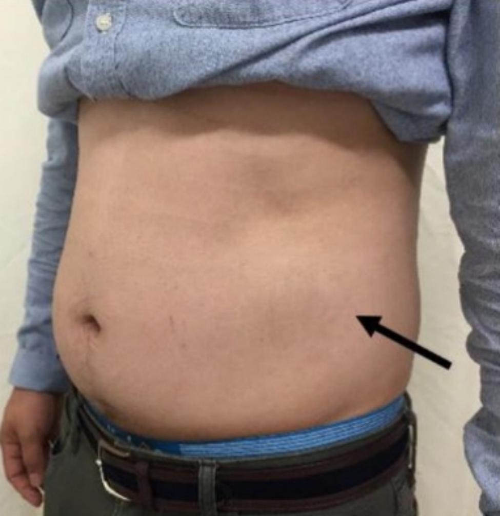
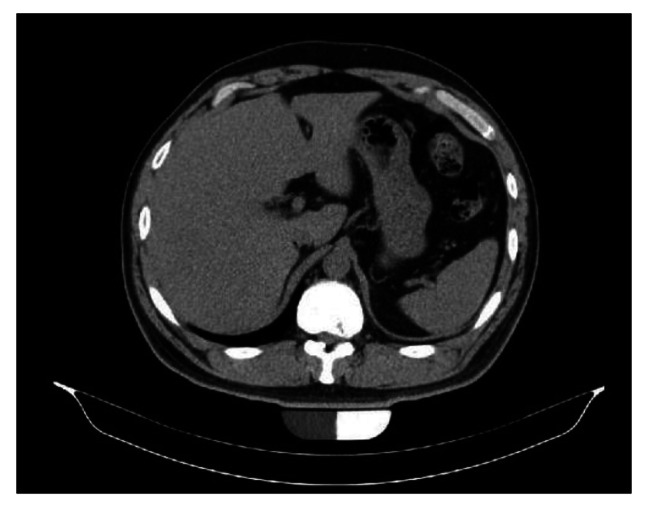
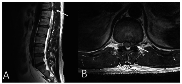
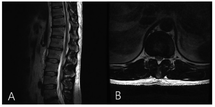
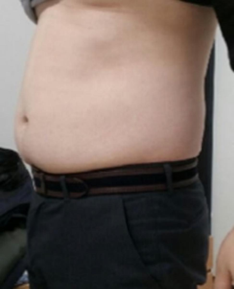
Case presentation: A 35-year-old man with no underlying disease or traumatic event presented with chief complaints of left flank pain and a protruding left lower abdominal mass that had formed one day earlier. There was no true abdominal hernia on abdominal computed tomography (CT), although CT and magnetic resonance imaging (MRI) showed a herniated soft (non-calcified) disc into the left neural foramen at the T11-12 level. A nonsteroidal anti-inflammatory drug was prescribed for the flank pain, and the patient was followed on a regular basis for six months. Follow-up MRI taken at the last visit showed complete resorption of the herniated disc. Abdominal pseudohernia and flank pain were also completely resolved.
Conclusion: We report a rare case of monoradiculopathy-induced abdominal pseudohernia caused by foraminal soft disc herniation at the T11-12 level. In patients who have an abdominal pseudohernia without herpes zoster infection, diabetes mellitus, or traumatic events, lower thoracic disc herniations should be included in differential diagnosis.
Keywords: Abdominal pseudohernia; Case report; Literature review; Thoracic disc herniation.
© 2023. The Author(s).
Conflict of interest statement
The authors declare no competing interests.
Figures
Similar articles
-
Abdominal wall pseudohernia - One secondary to a thoracic extraforaminal disc herniation and other due to thoracic paracentral disc protrusion.J Clin Orthop Trauma. 2022 May 16;30:101897. doi: 10.1016/j.jcot.2022.101897. eCollection 2022 Jul. J Clin Orthop Trauma. 2022. PMID: 35646588 Free PMC article.
-
Flank pseudohernia following posterior rib fracture: a case report.J Med Case Rep. 2016 Oct 1;10(1):273. doi: 10.1186/s13256-016-1054-9. J Med Case Rep. 2016. PMID: 27716425 Free PMC article. Review.
-
Symptoms of thoracolumbar junction disc herniation.Spine (Phila Pa 1976). 2001 Nov 15;26(22):E512-8. doi: 10.1097/00007632-200111150-00021. Spine (Phila Pa 1976). 2001. PMID: 11707722
-
Abdominal pseudohernia as an exceptional complication of herpes-zoster.Int J Surg Case Rep. 2024 Jan;114:109191. doi: 10.1016/j.ijscr.2023.109191. Epub 2023 Dec 24. Int J Surg Case Rep. 2024. PMID: 38150997 Free PMC article.
-
Intercostal lung herniation; a rare complication after mini-transthoracic approach (TTA) for thoracic disc herniation. Two case reports and review of literature.Eur Spine J. 2022 Dec;31(12):3708-3712. doi: 10.1007/s00586-021-07023-8. Epub 2022 Mar 23. Eur Spine J. 2022. PMID: 35318533 Review.
Cited by
-
Abdominal Pseudohernia Caused by Zoster Sine Herpete.Cureus. 2024 Nov 15;16(11):e73728. doi: 10.7759/cureus.73728. eCollection 2024 Nov. Cureus. 2024. PMID: 39677154 Free PMC article.
-
Abdominal pseudohernia caused by thoracic disk herniation: case series and review of the literature.J Surg Case Rep. 2025 Jan 23;2025(1):rjae822. doi: 10.1093/jscr/rjae822. eCollection 2025 Jan. J Surg Case Rep. 2025. PMID: 39850618 Free PMC article.
-
Spontaneous Resolution of Abdominal Pseudohernia Following Lung Cancer Surgery: A Case Report.Cureus. 2024 Jul 10;16(7):e64250. doi: 10.7759/cureus.64250. eCollection 2024 Jul. Cureus. 2024. PMID: 39130975 Free PMC article.
-
Abdominal Bulging Due to Abdominal Muscle Palsy Secondary to Herpes Zoster: A Report of a Rare Case.Cureus. 2024 Dec 26;16(12):e76440. doi: 10.7759/cureus.76440. eCollection 2024 Dec. Cureus. 2024. PMID: 39867103 Free PMC article.
References
Publication types
MeSH terms
LinkOut - more resources
Full Text Sources
Medical
