. 2023 Jun 28;51(3):1377-1385.
doi: 10.1042/BST20230121.

Caveolae and the oxidative stress response

Affiliations

Caveolae and the oxidative stress response

Yeping Wu et al. Biochem Soc Trans. .

Abstract

Oxidative stress is a feature of many disease conditions. Oxidative stress can activate a number of cellular pathways leading to cell death, including a distinct iron-dependent pathway involving lipid peroxidation, termed ferroptosis, but cells have evolved complex mechanisms to respond to these stresses. Here, we briefly summarise current evidence linking caveolae to the cellular oxidative stress response. We discuss recent studies in cultured cells and in an in vivo model suggesting that lipid peroxidation driven by oxidative stress causes disassembly of caveolae to release caveola proteins into the cell where they regulate the master transcriptional redox controller, nuclear factor erythroid 2-related factor 2. These studies suggest that caveolae maintain cellular susceptibility to oxidative stress-induced cell death and suggest a crucial role in cellular homeostasis and the response to wounding.

Keywords: Cavin1; NRF2; caveolae; cell death; lipid peroxidation; oxidative stress.


Conflict of interest statement

The authors declare that there are no competing interests associated with the manuscript.

Figures
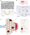
Figure 1.
Figure 1.. The role of caveolae in cellular response to oxidative stress.
(A) Top inset: caveolae as viewed by electron microscopy. Scale bar = 100 nm. Bottom inset: An illustration of caveolar structure in a cell under steady state conditions. (B) A diagram illustrating caveola-mediated cellular pathway in response to oxidative stress. Exogenous H2O2 stimulation, or GPX4 inactivation by ferroptosis inducer RSL3 [13,87], promotes ROS accumulation in cells. Excessive ROS-driven lipid peroxidation causes membrane damage and caveolar disassembly, leading to the release of Cavin1. This allows Cavin1 to target NRF2 in the cytosol and promote NRF2 ubiquitination (i), and NRF2 proteasomal degradation (ii). The binding of Cavin1 to NRF2 also inhibits the nuclear import of NRF2 (iii) [78]. The inhibition of NRF2 by Cavin1 through multiple mechanisms reduce the transcriptional levels of NRF2 downstream genes involved in oxidant defence, ultimately promoting ROS-driven cell death including ferroptosis and apoptosis. (C) A model for the role of caveolae in cellular oxidative stress response and its bearing in a physiological context of wound healing in the zebrafish. Upon tissue injury (a.), a paracrinal wave of ROS (represented by red arrows) such as H2O2 signals occur at the local wounding site [78,88]. Cells at steady state near the wound front experience oxidative stress which may trigger ROS-driven cell death (represented by b. and c.; dotted red arrow represents conversion of the state of a cell undergoing oxidative stress to a cell experiencing cell death). Regeneration pathways are subsequently triggered by cell death [79] (blue arrow) or ROS (d.) [3,89,90].

Similar articles

Cited by

References

    1. Hayes, J.D., Dinkova-Kostova, A.T. and Tew, K.D. (2020) Oxidative stress in cancer. Cancer Cell 38, 167–197 10.1016/j.ccell.2020.06.001 - DOI - PMC - PubMed
    1. Azzam, E.I., Jay-Gerin, J.P. and Pain, D. (2012) Ionizing radiation-induced metabolic oxidative stress and prolonged cell injury. Cancer Lett. 327, 48–60 10.1016/j.canlet.2011.12.012 - DOI - PMC - PubMed
    1. Dunnill, C., Patton, T., Brennan, J., Barrett, J., Dryden, M., Cooke, J.et al. (2017) Reactive oxygen species (ROS) and wound healing: the functional role of ROS and emerging ROS-modulating technologies for augmentation of the healing process. Int. Wound J. 14, 89–96 10.1111/iwj.12557 - DOI - PMC - PubMed
    1. Murphy, M.P., Bayir, H., Belousov, V., Chang, C.J., Davies, K.J.A., Davies, M.J., et al. (2022) Guidelines for measuring reactive oxygen species and oxidative damage in cells and in vivo. Nat. Metab. 4, 651–662 10.1038/s42255-022-00591-z - DOI - PMC - PubMed
    1. Conrad, M., Kagan, V.E., Bayir, H., Pagnussat, G.C., Head, B., Traber, M.G.et al. (2018) Regulation of lipid peroxidation and ferroptosis in diverse species. Genes Dev. 32, 602–619 10.1101/gad.314674.118 - DOI - PMC - PubMed

Publication types

Substances