. 2023 Jun 15;136(12):jcs260104.
doi: 10.1242/jcs.260104. Epub 2023 Jun 14.

Vinculin Y822 is an important determinant of ligand binding

Affiliations

Vinculin Y822 is an important determinant of ligand binding

Gillian DeWane et al. J Cell Sci. .

Abstract

Vinculin is an actin-binding protein present at cell-matrix and cell-cell adhesions, which plays a critical role in bearing force experienced by cells and dissipating it onto the cytoskeleton. Recently, we identified a key tyrosine residue, Y822, whose phosphorylation plays a critical role in force transmission at cell-cell adhesions. The role of Y822 in human cancer remains unknown, even though Y822 is mutated to Y822C in uterine cancers. Here, we investigated the effect of this amino acid substitution and that of a phosphodeficient Y822F vinculin in cancer cells. We observed that the presence of the Y822C mutation led to cells that proliferate and migrate more rapidly and contained smaller focal adhesions when compared to cells with wild-type vinculin. In contrast, the presence of the Y822F mutation led to highly spread cells with larger focal adhesions and increased contractility. Furthermore, we provide evidence that Y822C vinculin forms a disulfide bond with paxillin, accounting for some of the elevated phosphorylated paxillin recruitment. Taken together, these data suggest that vinculin Y822 modulates the recruitment of ligands.

Keywords: Cell–cell adhesion; Cell–matrix adhesion; Mechanotransduction; Vinculin.


Conflict of interest statement

Competing interests The authors declare no competing or financial interests.

Figures

Fig. 1.
Fig. 1.
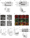
4T1 cells lacking vinculin have altered phenotypes. (A) Vinculin expression was stably inhibited using CRISPR. Two guide RNAs targeting mouse vinculin were designed and inserted into a plasmid containing Cas9. 4T1 cells were infected with the guide RNA-Cas9 plasmids, and two vinculin knockout clones were obtained (KO vin cl.1 and KO vin cl.2). Vinculin expression was examined by immunoblotting whole cell lysates with an antibody against vinculin or β-actin as a loading control. The graph beneath the immunoblots depicts vinculin levels normalized to vinculin levels in the parental cells; the data are mean±s.e.m., n=3 biologically independent samples. (B) The vinculin-knockout cells were smaller and more rounded when compared to 4T1 parental cells. Representative phase contrast images of the cells are shown from n=3 experimental repeats. Scale bars: 100 µm. (C) Immunofluorescence revealed a lack of detectable vinculin in knockout cells. Parental or vinculin knockout cells were fixed and stained with antibodies against phalloidin and vinculin; actin and vinculin were examined by confocal microscopy. Representative images from n=3 experimental repeats are shown. Scale bars: 10 µm. (D) Protein expression is decreased in vinculin-knockout cells. Talin, E-cadherin and paxillin expression was examined by immunoblotting whole-cell lysates; β-actin was used as a loading control. The graphs beneath the blots show the amount of each protein normalized to actin. The data are mean±s.e.m., n=3 biologically independent samples. *P<0.05 (two-tailed unpaired Student's t-test).
Fig. 2.
Fig. 2.
Y822C and Y822F vinculin expressing cells have different focal adhesion morphologies. (A) 4T1 vinculin-knockout cells were rescued with GFP-tagged versions of wild-type (WT) vinculin, Y822C or Y822F vinculin, or a GFP-only control. The expression of vinculin and GFP was examined by immunoblotting whole-cell lysates with an antibody against vinculin, GFP or β-actin as a loading control. The graph shows vinculin expression relative to total vinculin levels in the wild-type cells; data are mean±s.e.m., n=3 biologically independent samples. (B) The phosphorylation state of Y822 was examined in the cells. Lysates from the indicated cell lines were immunoblotted with antibodies against phosphorylated Y822 (p-Y822) then stripped and re-probed with antibodies that recognize total vinculin levels. The graph shows p-Y822 levels relative to total vinculin levels; data are mean±s.e.m., n=3 biologically independent samples. (C) The morphologies of the Y822C- and Y822F-expressing cells are altered. Representative phase contrast images of indicated cells from n=3 experimental repeats. Scale bars: 50 µm. (D–G) The focal adhesion phenotypes of Y822C and Y822F cells are different. (D) The indicated cells were fixed and stained with antibodies against phalloidin, and phalloidin and GFP expression was examined by confocal microscopy. Representative images are shown. Scale bars: 10 µm. (E–G) Cell area and focal adhesions containing GFP–vinculin were measured in ImageJ. (E) The cell area in the Y822F cells was larger compared to wild-type, Y822C and GFP-only cells. The graph shows total cell area, where the area of 30 cells was measured. n=3 biologically independent samples. (F) Y822C and Y822F vinculin-expressing cells showed an increased number of focal adhesions per cell compared to wild-type vinculin-expressing cells. The graph shows focal adhesion number per cell where focal adhesions in 45 cells were counted. n=3 biologically independent samples. (G) The focal adhesions in the Y822F cells were longer compared to wild-type cells, whereas the focal adhesions were smaller in the Y822C cells. The graph shows focal adhesion length where 665 focal adhesions were measured in 28 cells. n=4 biologically independent samples. Error bars in E–G are mean±s.e.m. (H) Myosin light chain phosphorylation (p-MLC) was elevated in Y822F cells. The cells were lysed and immunoblotted with antibodies that recognize phosphorylated MLC and then stripped and re-probed with antibodies that recognize total MLC levels. Results are mean±s.e.m.; n=3 biologically independent samples. *P<0.05 (two-tailed unpaired Student's t-test).
Fig. 3.
Fig. 3.
Y822C and Y822F vinculin-expressing cells have increased tumorigenicity. (A) 4T1 Y822F vinculin-expressing cells proliferated more than wild-type (WT) vinculin-expressing cells. Cells expressing the indicated GFP proteins were seeded into 96-well plates. After 72 h, cell viability was determined by means of an MTT assay. Results are mean±s.e.m.; n=5 biologically independent samples. (B–F) Y822C and Y822F cells proliferated more than wild-type vinculin cells in soft agar. (B) The indicated cells were seeded in 0.35% agarose and plated over a solidified 0.5% agarose bottom layer. The cells were incubated for 4 weeks. Representative images are shown; n=3 biologically independent samples. Scale bars: 1 mm. (C) Y822C cells grew more colonies than wild-type cells. Colony number was counted in ImageJ. n=3 biologically independent samples. (D–F) Y822C and Y822F colony size was different compared to wild-type cells. Colony size was counted with the Multi-point tool in ImageJ using three differently sized circles corresponding with small, medium and large sized colonies. (D,E) Y822C cells grew more small and medium colonies, respectively, whereas Y822F vinculin expressing cells grew more large colonies compared to wildtype cells (F). Results are mean±s.e.m.; n=3 biologically independent experiments. *P<0.05 (two-tailed unpaired Student's t-test). (G) representative images from wound healing assay for indicated cells; the edges of the wound are marked. Y822C and Y822F cells migrated faster than wild-type cells. Scale bars: 100 μm. (H) Y822F cells migrated to close a wound at a significantly faster rate than wild-type vinculin cells starting after 12 h of migration (significance denoted by solid black line). Y822C cells migrated to close the wound significantly faster than wild-type vinculin cells starting after 5 h of migration (significance denoted by dashed black line). Results are mean±s.e.m.; n=3 biologically independent experiments. *P<0.05 (two-way ANOVA with post-hoc Tukey's multiple comparison test).
Fig. 4.
Fig. 4.
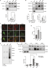
Y822C and Y822F vinculin mutants bind differently to binding partners and the cysteine at Y822C vinculin is necessary for this effect. (A) Y822F bound talin better than wild-type (WT) vinculin; Y822C bound talin less well. GFP or the GFP-tagged vinculins were immunoprecipitated, and the co-precipitating levels of talin were examined (IP). Average talin binding is shown relative to GFP or GFP–vinculin levels. Data are mean±s.e.m., n=3 biologically independent samples. (B) Y822C vinculin co-precipitated with more paxillin but less β-actin compared to wild-type vinculin. Y822F vinculin co-precipitated with more paxillin, but similar amounts of β-actin compared to wild-type vinculin. GFP or GFP-tagged vinculin was immunoprecipitated from 4T1 cells, and the co-precipitating levels of paxillin and β-actin were examined (IP). Average paxillin or β-actin binding is shown relative to GFP or GFP-tagged vinculin levels. Data are mean±s.e.m., n=3 biologically independent samples. (C,D) More phosphorylated paxillin colocalized with Y822C vinculin. (C) The indicated cell lines were stained with antibodies against phosphorylated paxillin (p-Paxillin) and visualized using confocal microscopy. Representative images are shown. Scale bars: 10 µm. (D) The graphs depict a R value for the colocalization of phospho-paxillin and vinculin. The analysis revealed more phosphorylated paxillin colocalized with Y822C vinculin-containing focal adhesions. Cells from three separate field of views were chosen. Data are mean±s.e.m., n=3 biologically independent samples. *P<0.05 (ordinary one-way ANOVA and Dunnett's multiple comparisons tests). (E) The association of vinculin mutants with paxillin under non-reducing conditions. GFP-tagged vinculins were immunoprecipitated (IP:GFP) from cells, run on a gel under non-reducing conditions, and then probed for the presence of paxillin or GFP-vinculin (IB: Paxillin). The graph depicts the amount of paxillin recovered as a function of GFP–vinculin levels. Data are mean±s.e.m., n=3 biologically independent samples. (F) Pre-incubation of cells with the antioxidant NAC decreased paxillin binding to Y822C vinculin in 4T1 cells and does not significantly change paxillin co-precipitation with wild-type or Y822F vinculin. Cells were pre-treated with 10 mM NAC for 1 h then GFP or GFP–vinculin was immunoprecipitated, and the co-precipitating levels of paxillin were examined. Average paxillin binding is shown relative to GFP or GFP–vinculin levels. Data are mean±s.e.m., n=3 biologically independent samples. *P<0.05 (two-tailed unpaired Student's t-test).
Fig. 5.
Fig. 5.
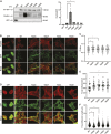
A serine or alanine substitution at Y822 does not affect paxillin binding or focal adhesion length. 4T1 cells stably expressing the indicated vinculin variants were generated. (A–C) The Y822A or Y822S mutations abrogated the effect of Y822C on paxillin binding and co-localization with vinculin. WT, wild-type. (A) GFP or GFP-tagged vinculin was immunoprecipitated, and the co-precipitating levels of paxillin were examined. Average paxillin binding is shown relative to GFP or GFP-vinculin levels. Data are mean±s.e.m., n=3 biologically independent samples. (B,C) Cells were stained with antibodies against paxillin and the colocalization of paxillin with the GFP proteins was examined. Representative images are shown (B). Scale bars: 10 µm. The graph in C represents a quantification of the amount of paxillin as a function of vinculin in the focal adhesions revealed more paxillin colocalized with Y822C- and Y822F-vinculin-containing focal adhesions. Graphs indicating intensity show fluorescence intensity of the ROI of paxillin within the focal adhesion. Data are mean for n=3 biologically independent samples. *P<0.05 (ordinary one-way ANOVA and Dunnett's multiple comparisons tests). (D–F) Y822A and Y822S cells had focal adhesion phenotypes similar to those in wild-type cells. (D) GFP or GFP-tagged vinculin-expressing cells were fixed and stained with antibodies against phalloidin; GFP expression was examined by confocal microscopy. Representative images are shown. Scale bars: 10 µm. (E) Y822C, Y822F, Y822A and Y822S vinculin cells showed increased number of focal adhesions per cell compared to cells expressing wild-type vinculin. Focal adhesions containing GFP–vinculin were counted in ImageJ. Graph shows focal adhesion number per cell where focal adhesions in 45 cells were counted. Data are mean±s.e.m., n=3 biologically independent samples. (F) Y822A and Y822S vinculin cells had similarly sized focal adhesions to those in wild-type cells. Focal adhesions containing GFP–vinculin were measured in ImageJ. The graph shows focal adhesion length where 666 focal adhesions were measured in 27 cells. n=3 biologically independent samples. *P<0.05 (two-tailed unpaired Student's t-test).

References

    1. Abé, C., Dietrich, F., Gajula, P., Benz, M., Vogel, K.-P., Van Gastel, M., Illenberger, S., Ziegler, W. H. and Steinhoff, H.-J. (2011). Monomeric and dimeric conformation of the vinculin tail five-helix bundle in solution studied by EPR spectroscopy. Biophys. J. 101, 1772-1780. 10.1016/j.bpj.2011.08.048 - DOI - PMC - PubMed
    1. Auernheimer, V. and Goldmann, W. H. (2014). Serine phosphorylation on position 1033 of vinculin impacts cellular mechanics. Biochem. Biophys. Res. Commun. 450, 1095-1098. 10.1016/j.bbrc.2014.06.122 - DOI - PubMed
    1. Auernheimer, V., Lautscham, L. A., Leidenberger, M., Friedrich, O., Kappes, B., Fabry, B. and Goldmann, W. H. (2015). Vinculin phosphorylation at residues Y100 and Y1065 is required for cellular force transmission. J. Cell Sci. 128, 3435-3443. 10.1242/jcs.172031 - DOI - PMC - PubMed
    1. Bakolitsa, C., Cohen, D. M., Bankston, L. A., Bobkov, A. A., Cadwell, G. W., Jennings, L., Critchley, D. R., Craig, S. W. and Liddington, R. C. (2004). Structural basis for vinculin activation at sites of cell adhesion. Nature 430, 583-586. 10.1038/nature02610 - DOI - PubMed
    1. Ballestrem, C., Erez, N., Kirchner, J., Kam, Z., Bershadsky, A. and Geiger, B. (2006). Molecular mapping of tyrosine-phosphorylated proteins in focal adhesions using fluorescence resonance energy transfer. J. Cell Sci. 119, 866-875. 10.1242/jcs.02794 - DOI - PubMed

Publication types