. 2023 Sep;50(9):5875-5883.
doi: 10.1002/mp.16474. Epub 2023 May 30.

Characterization of a diode dosimeter for UHDR FLASH radiotherapy

Affiliations

Characterization of a diode dosimeter for UHDR FLASH radiotherapy

Mahbubur Rahman et al. Med Phys. 2023 Sep.

Abstract

Background: Ultra-high dose rate (UHDR) FLASH beams typically deliver dose at rates of >40 Gy/sec. Characterization of these beams with respect to dose, mean dose rate, and dose per pulse requires dosimeters which exhibit high temporal resolution and fast readout capabilities.

Purpose: A diode EDGE Detector with a newly designed electrometer has been characterized for use in an UHDR electron beam and demonstrated appropriateness for UHDR FLASH radiotherapy dosimetry.

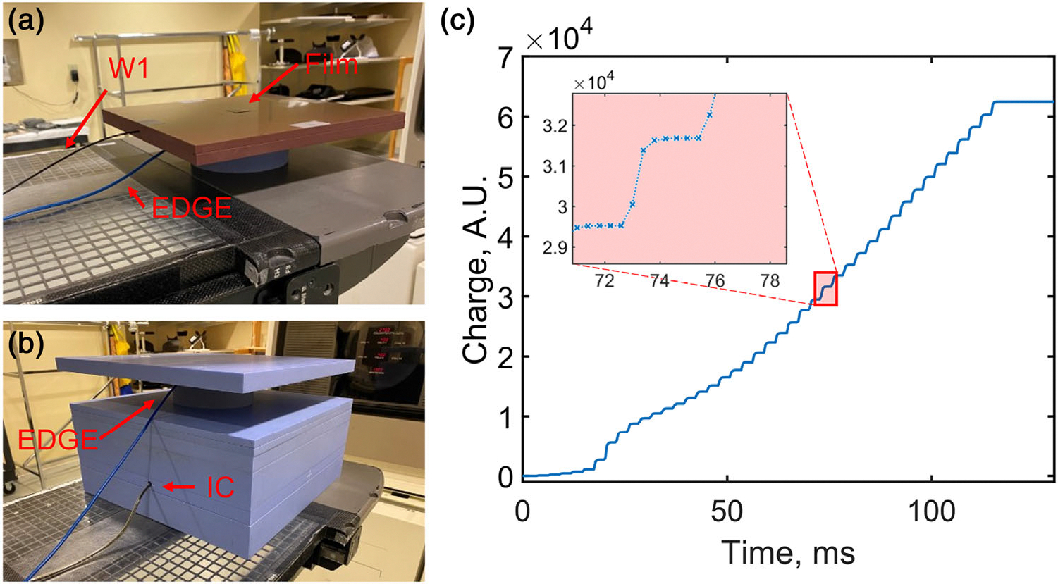
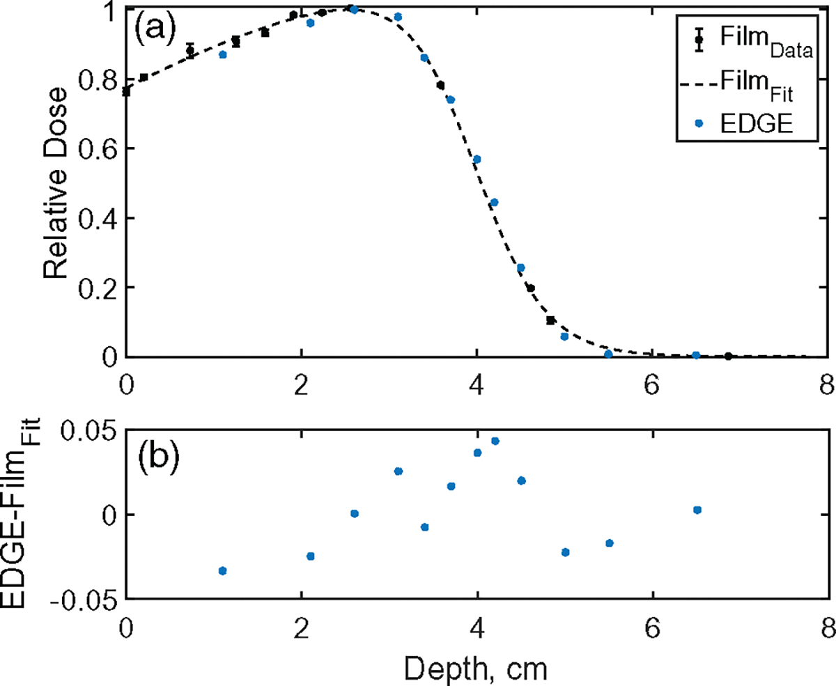
Methods: Dose linearity, mean dose rate, and dose per pulse dependencies of the EDGE Detector were quantified and compared with dosimeters including a W1 scintillator detector, radiochromic film, and ionization chamber that were irradiated with a 10 MeV UHDR beam. The dose, dose rate, and dose per pulse were controlled via an in-house developed scintillation-based feedback mechanism, repetition rate of the linear accelerator, and source-to-surface distance, respectively. Depth-dose profiles and temporal profiles at individual pulse resolution were compared to the film and scintillation measurements, respectively. The radiation-induced change in response sensitivity was quantified via irradiation of ∼5kGy.

Results: The EDGE Detector agreed with film measurements in the measured range with varying dose (up to 70 Gy), dose rate (nearly 200 Gy/s), and dose per pulse (up to 0.63 Gy/pulse) on average to within 2%, 5%, and 1%, respectively. The detector also agreed with W1 scintillation detector on average to within 2% for dose per pulse (up to 0.78 Gy/pulse). The EDGE Detector signal was proportional to ion chamber (IC) measured dose, and mean dose rate in the bremsstrahlung tail to within 0.4% and 0.2% respectively. The EDGE Detector measured percent depth dose (PDD) agreed with film to within 3% and per pulse output agreed with W1 scintillator to within -6% to +5%. The radiation-induced response decrease was 0.4% per kGy.

Conclusions: The EDGE Detector demonstrated dose linearity, mean dose rate independence, and dose per pulse independence for UHDR electron beams. It can quantify the beam spatially, and temporally at sub millisecond resolution. It's robustness and individual pulse detectability of treatment deliveries can potentially lead to its implementation for in vivo FLASH dosimetry, and dose monitoring.

Keywords: EDGE detector; FLASH; diode; film; ionization chamber; ultra-high dose rate.


Conflict of interest statement

CONFLICT OF INTEREST STATEMENT

The authors declare no conflicts of interest.

Figures

FIGURE 1
FIGURE 1
(a) Setup for data acquisition from both the W1 detector and characterization of the EDGE Detector. Film on top of the stack of solid water phantom was used for calibration of the W1 detector and verifying dose for each delivery. (b) Setup for data acquisition from both ion chamber (IC) in the Bremsstrahlung tail (17 cm depth) and EDGE Detector (both calibrated to dose measured at 2 cm depth from film. (c) Example Snapshot data acquired from the EDGE Detector with a zoomed in plot of charge accumulation from an example individual pulse at 0.4 ms sampling rate. Data acquisition is not synchronous to pulse timing. In the zoomed in plot, the pulse occurred approximately 0.06 ms prior to the measurement at 73 ms, which fits the exponential charge growth at points 73.0 through 75.0 ms with a 200 μs time constant.
FIGURE 2
FIGURE 2
W1 and EDGE Detector response in comparison to Gafchromic film and ion chamber (IC) with varying dose. The intended dose delivery was controlled based on W1 detector response. Dose was calculated from integrated total dose delivery/number of pulses.
FIGURE 3
FIGURE 3
W1 and EDGE Detector response in comparison to Gafchromic film and EDGE detector response in comparison to ion chamber (IC) for varying mean dose rate. The mean dose rate was varied by changes in the repetition rate of the pulses delivered by the LINAC. The mean dose rate values were calculated from integrated total dose delivery/number of pulses.
FIGURE 4
FIGURE 4
(a) W1 and EDGE Detector dose per pulse response comparison to Gafchromic film with varying SSD by adjusting the couch’s vertical position. The dose per pulse was varied by changing the source-to-surface distance and adjusting the couch’s vertical position. Dose per pulse values were calculated from integrated total dose delivery/number of pulses. (b) EDGE detector comparison to W1 for individual pulses from 3 deliveries at each of 100 cm SSD position. The varying in individual pulses can be attributed to the LINAC’s ramp up period.
FIGURE 5
FIGURE 5
(a) Percent depth dose curve (PDD) measured by film and EDGE Detector for the 10 MeV UHDR electron beam. Normalized EDGE and film dose values were calculated from integrated total dose delivery/number of pulses. EDGE detector measurement depths ranged from 1.15 to 6.55 cm. (b) Difference between PDD fitted to Film profile and EDGE Detector measurements.
FIGURE 6
FIGURE 6
(a) Relative per pulse beam output from the LINAC delivering UHDR electron beams measured by both the W1 and the EDGE Detector. (b) Relative ratio between EDGE detector and W1 scintillator response.
FIGURE 7
FIGURE 7
Long-term sensitivity of the EDGE Detector with respect to the response of the ion chamber (IC).

References

    1. Alaghband Y, Cheeks SN, Allen BD, et al. Neuroprotection of radiosensitive juvenile mice by ultra-high dose rate FLASH irradiation. Cancers. 2020;12(6):1671. doi:10.3390/cancers12061671 - DOI - PMC - PubMed
    1. Vozenin MC, Hendry JH, Limoli CL. Biological benefits of ultra-high dose rate FLASH radiotherapy: sleeping beauty awoken. Clin Oncol. 2019;31(7):407–415. doi:10.1016/j.clon.2019.04.001 - DOI - PMC - PubMed
    1. Favaudon V, Caplier L, Monceau V, et al. Ultrahigh dose-rate FLASH irradiation increases the differential response between normal and tumor tissue in mice. Sci Transl Med. 2014;6(245):245ra93–245ra93.doi:10.1126/scitranslmed.3008973 - DOI - PubMed
    1. Diffenderfer ES, Verginadis II, Kim MM, et al. Design, implementation, and in vivo validation of a novel proton FLASH radiation therapy system. Int J Radiat Oncol Biol Phys. 2020;106(2):440–448. doi:10.1016/j.ijrobp.2019.10.049 - DOI - PMC - PubMed
    1. Fouillade C, Curras-Alonso S, Giuranno L, et al. FLASH irradiation spares lung progenitor cells and limits the incidence of radio-induced senescence. Clin Cancer Res. 2020;26(6):1497–1506. doi:10.1158/1078-0432.CCR-19-1440 - DOI - PubMed

LinkOut - more resources