. 2023 Oct;20(8):3191-3203.
doi: 10.1111/iwj.14198. Epub 2023 May 30.

Identification and verification of ferroptosis-related genes in diabetic foot using bioinformatics analysis

Affiliations

Identification and verification of ferroptosis-related genes in diabetic foot using bioinformatics analysis

Xiaoxiang Wang et al. Int Wound J. 2023 Oct.

Abstract

Ferroptosis is a novel form of cell death that plays a key role in several diseases, including inflammation and tumours; however, the role of ferroptosis-related genes in diabetic foot remains unclear. Herein, diabetic foot-related genes were downloaded from the Gene Expression Omnibus and the ferroptosis database (FerrDb). The least absolute shrinkage and selection operator regression algorithm was used to construct a related risk model, and differentially expressed genes were analysed through immune infiltration. Finally, we identified relevant core genes through a protein-protein interaction network, subsequently verified using immunohistochemistry. Comprehensive analysis showed 198 genes that were differentially expressed during ferroptosis. Based on functional enrichment analysis, these genes were primarily involved in cell response, chemical stimulation, and autophagy. Using the CIBERSORT algorithm, we calculated the immune infiltration of 22 different types of immune cells in diabetic foot and normal tissues. The protein-protein interaction network identified the hub gene TP53, and according to immunohistochemistry, the expression of TP53 was high in diabetic foot tissues but low in normal tissues. Accordingly, we identified the ferroptosis-related gene TP53 in the diabetic foot, which may play a key role in the pathogenesis of diabetic foot and could be used as a potential biomarker.

Keywords: TP53; bioinformatics analysis; diabetic foot; ferroptosis; inflammatory reaction.


Conflict of interest statement

The authors declare that there is no conflict of interest.

Figures
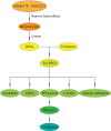
FIGURE 1
FIGURE 1
Study design and workflow.
FIGURE 2
FIGURE 2
Dataset de‐batching effect. (A) Gene expression level statistics of the dataset before de‐batching. (B) Gene expression level statistics of the dataset after de‐batching. (C) Density distribution of dataset before de‐batching. (D) Density distribution of dataset after de‐batch©. (E) UMAP distribution of dataset before de‐batching. (F) UMAP distribution of dataset after de‐batching. UMAP, Uniform Manifold Approximation and Projection.
FIGURE 3
FIGURE 3
Differentially expressed genes between patients with diabetic foot and normal control group. (A) Volcanic map of DEGs in diabetic foot and control group in GSE80178 and GSE29221 datasets. Red nodes indicate upregulated DEGs, blue nodes indicate downregulated DEGs and grey nodes indicate genes that are not significantly differentially expressed. (B) Heat map of expression levels of diabetic foot DEGs: red indicates disease samples, blue indicates normal control samples, pink indicates high gene expression and light blue indicates low gene expression. (C) Venn diagram shows 198 ferroptosis‐related DEGs. DEGs differentially expressed genes.
FIGURE 4
FIGURE 4
Enrichment analysis of Fer‐DEGs. (A) Biological process. (B) Cellular component. (C) Molecular function. (D) KEGG enrichment analysis. Fer‐DEGs, ferroptosis‐related differentially expressed genes; KEGG, Kyoto Encyclopedia of Genes and Genomes.
FIGURE 5
FIGURE 5
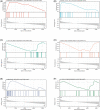
Gene set enrichment analysis. (A) Enrichment plot of “porphyrin and chlorophyll metabolism” with enrichment score 0.61. (B) Enrichment plot of “citrate cycle tca cycle” with enrichment score 0.48. (C) Enrichment plot of “N glycan biosynthesis” with enrichment score −0.60. (D) Enrichment plot of “ether lipid metabolism” with enrichment score −0.54. (E) Enrichment plot of “Parkinson's disease” with enrichment score 0.50. (E) Enrichment plot of “O‐glycan biosynthesis” with enrichment score −0.45.
FIGURE 6
FIGURE 6
Construction of diabetic foot model. (A, B) Screening of gene signatures from Fer‐DEGs using the LASSO algorithm. (C) Forest plot of gene signatures in patients with DM. (D) Receiver operating characteristic (ROC) curve of predicted risk scores in patients with DM. DM, diabetes mellitus; Fer‐DEGs, ferroptosis‐related differentially expressed genes; LASSO, least absolute shrinkage and selection operator.
FIGURE 7
FIGURE 7
CIBERSORT immune infiltration assessment. (A) Bar charts of 22 immune cell proportions in diabetic foot and control samples. The first 21 are diabetic feet, and the last 15 are controls. (B) Correlation heat map of 22 kinds of immune cells in diabetic foot samples. (*P < 0.05, **P < 0.01, ***P < 0.001, ****P < 0.0001, NSp≥0.05).
FIGURE 8
FIGURE 8
PPI network. (A) PPI network of Fer‐DEGs. (B) Red nodes indicate upregulated Fer‐DEGs and green nodes indicate downregulated Fer‐DEGs. (C) The MCODE plugin in Cytoscape was used to determine the module with the highest score. (D) Verification of Hub gene. Fer‐DEGs, ferroptosis‐related differentially expressed genes; PPI, protein–protein interaction.
FIGURE 9
FIGURE 9
Expression of the hub gene in normal skin and diabetic foot tissues. (A) Expression of TP53 in normal tissues. (B) Expression of TP53 in diabetic foot tissues.

Similar articles

Cited by

References

    1. Mayer‐Davis EJ, Lawrence JM, Dabelea D, et al. Incidence trends of type 1 and type 2 diabetes among youths, 2002‐2012. N Engl J Med. 2017;376(15):1419‐1429. - PMC - PubMed
    1. Yang L, Shao J, Bian Y, et al. Prevalence of type 2 diabetes mellitus among inland residents in China (2000‐2014): a meta‐analysis. J Diabetes Investig. 2016;7(6):845‐852. - PMC - PubMed
    1. Messenger G, Taha N, Sabau S, AlHubail A, Aldibbiat AM. Is there a role for informal caregivers in the Management of Diabetic Foot Ulcers? A Narrative Review. Diabetes Ther. 2019;10(6):2025‐2033. - PMC - PubMed
    1. Rayman G, Vas P, Dhatariya K, et al. Guidelines on use of interventions to enhance healing of chronic foot ulcers in diabetes (IWGDF 2019 update). Diabetes Metab Res Rev. 2020;36(Suppl 1):e3283. - PubMed
    1. Lin CJ, Lan YM, Ou MQ, Ji LQ, Lin SD. Expression of miR‐217 and HIF‐1alpha/VEGF pathway in patients with diabetic foot ulcer and its effect on angiogenesis of diabetic foot ulcer rats. J Endocrinol Investig. 2019;42(11):1307‐1317. - PubMed