. 2023 Jul;37(7):e22983.
doi: 10.1096/fj.202201868RR.

Aberrant activation of Notch1 signaling in the mouse uterine epithelium promotes hyper-proliferation by increasing estrogen sensitivity

Affiliations

Aberrant activation of Notch1 signaling in the mouse uterine epithelium promotes hyper-proliferation by increasing estrogen sensitivity

Qi-Xin Xu et al. FASEB J. 2023 Jul.

Abstract

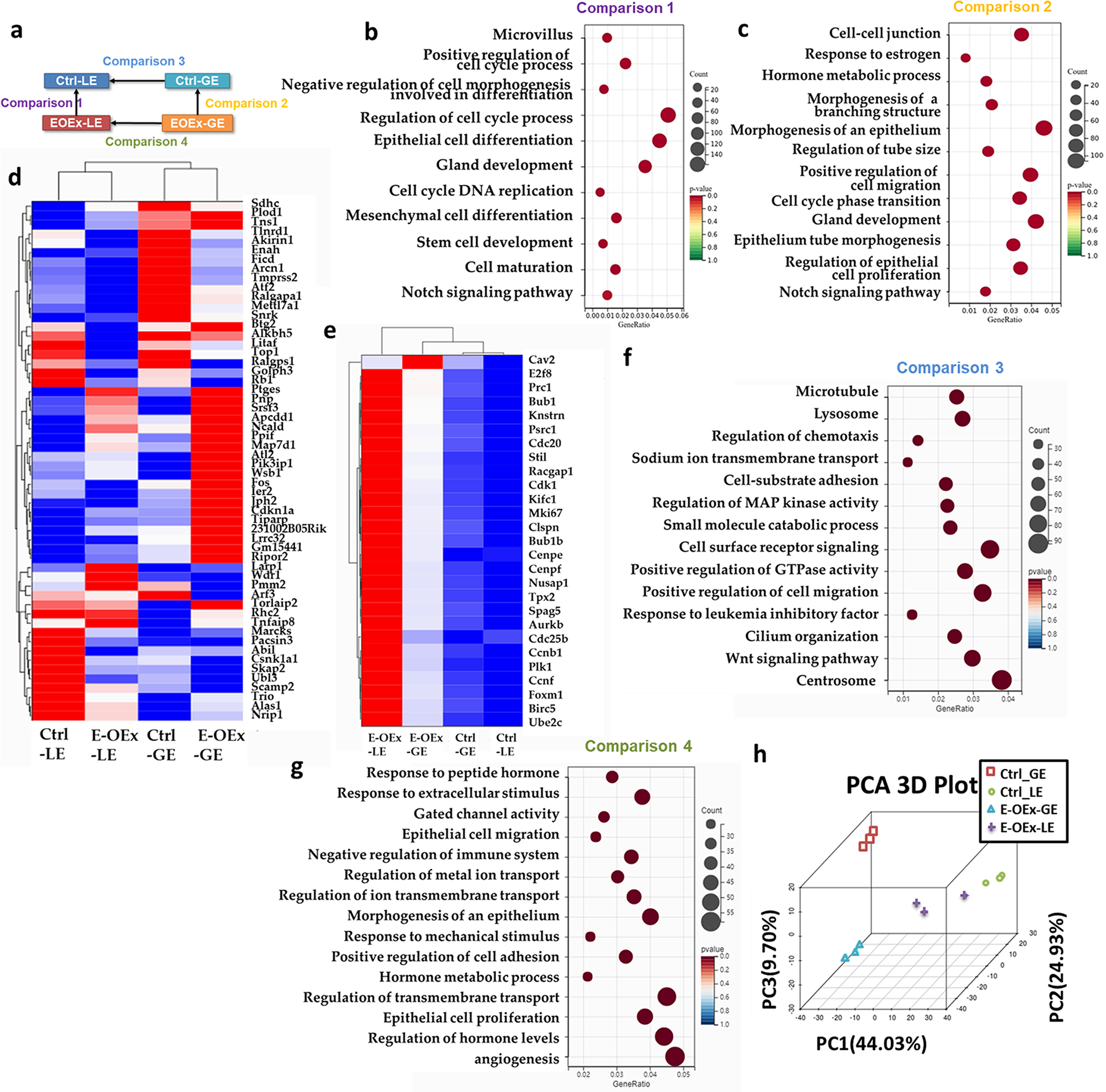
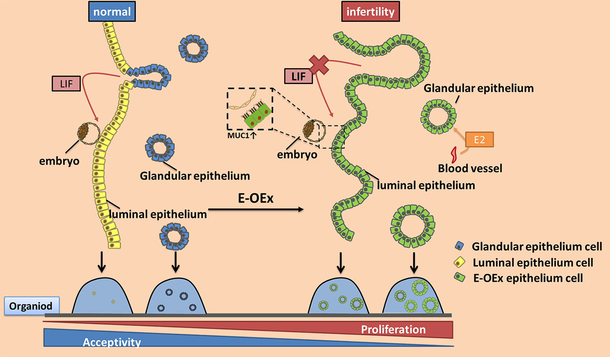
In mammals, the endometrium undergoes dynamic changes in response to estrogen and progesterone to prepare for blastocyst implantation. Two distinct types of endometrial epithelial cells, the luminal (LE) and glandular (GE) epithelial cells play different functional roles during this physiological process. Previously, we have reported that Notch signaling plays multiple roles in embryo implantation, decidualization, and postpartum repair. Here, using the uterine epithelial-specific Ltf-iCre, we showed that Notch1 signaling over-activation in the endometrial epithelium caused dysfunction of the epithelium during the estrous cycle, resulting in hyper-proliferation. During pregnancy, it further led to dysregulation of estrogen and progesterone signaling, resulting in infertility in these animals. Using 3D organoids, we showed that over-activation of Notch1 signaling increased the proliferative potential of both LE and GE cells and reduced the difference in transcription profiles between them, suggesting disrupted differentiation of the uterine epithelium. In addition, we demonstrated that both canonical and non-canonical Notch signaling contributed to the hyper-proliferation of GE cells, but only the non-canonical pathway was involved with estrogen sensitivity in the GE cells. These findings provided insights into the effects of Notch1 signaling on the proliferation, differentiation, and function of the uterine epithelium. This study demonstrated the important roles of Notch1 signaling in regulating hormone response and differentiation of endometrial epithelial cells and provides an opportunity for future studies in estrogen-dependent diseases, such as endometriosis.

Keywords: Notch1 signaling; estrogen sensitivity; glandular epithelium; hyper-proliferation; luminal epithelium.


Conflict of interest statement

Conflict of Interest

The authors declare no competing financial interests.

Figures

Fig. 1.
Fig. 1.
Defective uterine receptivity in E-OEx mice. (a) Generation strategy for conditional overexpression of N1ICD in the epithelial endometrium (Epithelium-specific overexpressed N1ICD, E-OEx, Ltfcre/+Rosa26N1ICD/+). (b) The mRNA expression of Notch1 (N1ICD), Egfp, Hey1, and Hes5 in the Ctrl (Ltf+/+Rosa26N1ICD/+) and E-OEx mice (n=3, t test). (c) Comparison of the protein expression of NOTCH1 in Ctrl and E-OEx mice. (d) Implantation sites are shown as blue dots in Ctrl, E-OEx, E-OEx mice supplemented with LIF at 3.5 dpc (E-OEx-Lif), and E-OEx mice with Rbpj knockout (Ltfcre/+Rosa26N1ICD/+RbpjF/F, E-OEx-KO) mice at 4.5 dpc. The black arrow marks the implantation sites. (e) The histological staining of implantation sites. The black arrow marks the implantation sites, the red arrow marks the floating embryos. (f) Morphology and number of blastocysts flushed out of one uterine horn of Ctrl and E-OEx mice on 3.5 dpc (n=5 in Ctrl mice, n=6 in E-OEx mice, t test). (g) The mRNA level of Lif in the uterus from Ctrl and E-OEx mice on 3.5 dpc (n=3, t test). (h) Weight of stimulated horns and non-stimulated uterine horns in Ctrl and E-OEx mice. (i) Uterine morphology in response to an artificial decidualization stimulus in Ctrl and E-OEx mice. Left horns served as non-stimulated controls. (j-l) qPCR analysis of decidua markers Wnt4, Bmp2, and Prl8a2 in stimulated and non-stimulated uterine horns in Ctrl and E-OEx mice (n=3, t test). Black dotted lines mark the LE, red arrow mark the GE. Values are mean ± SEM.
Fig. 2.
Fig. 2.
Overactivation of Notch1 signaling results in a dysregulated luminal epithelium via hormone-controlled cell proliferation. (a) H&E and Ki67 staining in the uterus of Ctrl, E-OEx, and E-OEx-KO mice on 3.5 dpc. Black dotted lines mark the LE, red arrow marks the GE, Blue font marks the location of Stroma (S). (b) 3D images of the uterine lumen of Ctrl and E-OEx females on 3.5 dpc. (c) The mRNA expression of progesterone (P4) target genes in the uterus of Ctrl and E-OEx mice on 3.5 dpc (n=3, t test). (d) The mRNA expression of and estrogen (E2) target genes in the uterus of Ctrl and E-OEx mice on 3.5 dpc (n=3, t test). (e) The immunostaining of ESR1, PR, and Ac-Tubulin in the uterus of Ctrl and E-OEx mice on 3.5 dpc. (f) Digital H-Score of ESR1 expression in luminal (LE) and glandular epithelium (GE) of Ctrl and E-OEx uterus shown in e. (g) Digital H-Score of PR expression in luminal (LE) and glandular epithelium (GE) of Ctrl and E-OEx uterus shown in e (n=3 for LE, n=9 for GE, Two-way ANOVA). Values are mean ± SEM. Scale bar = 100 μm
Fig. 3.
Fig. 3.
Effects of Notch1 signaling on proliferative potential of organoids derive from LE and GE. (a) Comparison of the isolated LE morphology of Ctrl and E-OEx mice. (b, c) mRNA expression of Spink3 and Foxa2 in LE, GE, and S isolated from Ctrl and E-OEx mice on 3.5 dpc (n>3, Two-way ANOVA). (d) Morphological and histological images of LE and GE- derived organoids from Ctrl and E-OEx mice following 3 days of culture. (e,f) The number (e) and diameter (f) of the LE and GE- derived organoids from Ctrl and E-OEx mice after 3 days of culture (n>3, Two-way ANOVA). (g) Quantification of the distribution of organoid sizes after 3 days in culture. (h) Morphological images of LE and GE- derived organoids after activation of N1ICD by adenovirus with Cre recombinase (Cre-Ad) following 5 days of culture. (i) Diameter of organoids in (h) after 5 days of culture (n>3, Two-way ANOVA). Values are mean ± SEM.
Fig. 4.
Fig. 4.
RNA-seq data analysis of LE and GE from Ctrl and E-OEx mice. (a) Diagram of the comparisons setup. (b) Gene ontology (GO) analysis of comparison 1. (c) Gene ontology (GO) analysis of comparison 2. (d) Clustered heatmap of estrogen-sensitive genes in Ctrl-LE, Ctrl-GE, E-OEx-LE, and E-OEx-GE. (e) Heatmap depicts the expression pattern proliferation associated genes in Ctrl-LE, Ctrl-GE, E-OEx-LE, and E-OEx-GE. (f) Gene ontology (GO) analysis of DEGs in comparison 3 but not 4. (g) Gene ontology (GO) analysis of DEGs in comparison 4 but not 3. (h) PCA analysis of Ctrl-LE, Ctrl-GE, E-OEx-LE, and E-OEx-GE.
Fig. 5.
Fig. 5.
The sensitized proliferation in response to E2 in the E-OEx mice. (a) Ki67 staining in the Ctrl and E-OEx uterus at different stages of the estrous cycle. (b) Digital H-Score of GE KI67 expression in a (n=12, Two-way ANOVA). (c) Digital H-Score of LE KI67 expression in a (n=3, Two-way ANOVA). (d) H&E staining of the uterus from ovariectomized Ctrl and E-OEx mice after being treated with sesame oil (Vehicle), lower (10 ng/0.1ml) and higher (100 ng/0.1 ml) E2, and E2 + P4 (100 ng/0.1 ml and 1 mg/0.1 ml). Black arrow showed the glands. Black dotted lines mark the LE, red arrow marks the GE, Blue fonts mark the location of Stroma (S). (e) Cell counts per gland in the uterus from Ctrl and E-OEx mice (n=12, Two-way ANOVA). Immunohistochemistry of the KI67 (f) and FOXA2 (g) of Ctrl, E-OEx, and E-OEx-KO uterus. (h) KI67 positive cells/total cell of GE in the uterus from Ctrl and E-OEx mice (n=12, Two-way ANOVA). (i) 3D imaging of the Ctrl and E-OEx uterus treated with Vehicle or 100ng/0.1ml E2, glands are stained with FOXA2. Values are mean ± SEM.
Fig. 6.
Fig. 6.
A diagrammatic summary of the results of this study. Over-activation of Notch1 signaling results in hyperproliferation of both LE and GE cells, weakens the differentiation features and function of GE, resulting in decreased LIF levels and impaired uterine receptivity. Moreover, the higher level of Notch1 signaling enhances the sensitivity of the GE to estrogen, which allows the GE cells to proliferate under lower estrogen levels during estrous cycle, and results in hyperproliferation of GE. Similarly, in the in vitro culture, over-activation of Notch1 signaling increases the regeneration capacity of both LE and GE in organoid.

References

    1. Kelleher AM, Demayo FJ and Spencer TE Uterine Glands: Developmental Biology and Functional Roles in Pregnancy. Endocr Rev. 2019; 40: 1424–1445. - PMC - PubMed
    1. Wood GA, Fata JE, Watson KLM and Khokha R Circulating hormones and estrous stage predict cellular and stromal remodeling in murine uterus. Reproduction. 2007; 133: 1035–1044. - PubMed
    1. Cha J, Sun X and Dey SK Mechanisms of implantation: strategies for successful pregnancy. Nat Med. 2012; 18: 1754–1767. - PMC - PubMed
    1. Habiba M, Heyn R, Bianchi P, Brosens I and Benagiano G The development of the human uterus: morphogenesis to menarche. Hum Reprod Update. 2021; 27: 1–26. - PubMed
    1. Feng D, Menger MD, Wang H and Laschke MW Luminal epithelium in endometrial fragments affects their vascularization, growth and morphological development into endometriosis-like lesions in mice. Dis Model Mech. 2014; 7: 225–232. - PMC - PubMed

Publication types

LinkOut - more resources