. 2023 Apr 27;26(5):106764.
doi: 10.1016/j.isci.2023.106764. eCollection 2023 May 19.

Involvement of Mrgprd-expressing nociceptors-recruited spinal mechanisms in nerve injury-induced mechanical allodynia

Affiliations

Involvement of Mrgprd-expressing nociceptors-recruited spinal mechanisms in nerve injury-induced mechanical allodynia

Liangbiao Wang et al. iScience. .

Abstract

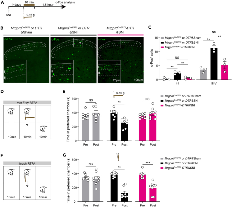
Mechanical allodynia and hyperalgesia are intractable symptoms lacking effective clinical treatments in patients with neuropathic pain. However, whether and how mechanically responsive non-peptidergic nociceptors are involved remains elusive. Here, we showed that von Frey-evoked static allodynia and aversion, along with mechanical hyperalgesia after spared nerve injury (SNI) were reduced by ablation of MrgprdCreERT2-marked neurons. Electrophysiological recordings revealed that SNI-opened Aβ-fiber inputs to laminae I-IIo and vIIi, as well as C-fiber inputs to vIIi, were all attenuated in Mrgprd-ablated mice. In addition, priming chemogenetic or optogenetic activation of Mrgprd+ neurons drove mechanical allodynia and aversion to low-threshold mechanical stimuli, along with mechanical hyperalgesia. Mechanistically, gated Aβ and C inputs to vIIi were opened, potentially via central sensitization by dampening potassium currents. Altogether, we uncovered the involvement of Mrgprd+ nociceptors in nerve injury-induced mechanical pain and dissected the underlying spinal mechanisms, thus providing insights into potential therapeutic targets for pain management.

Keywords: Cell biology; Cellular neuroscience; Neuroscience.


Conflict of interest statement

The authors declare no competing interests.

Figures

None
Graphical abstract
Figure 1
Figure 1
Attenuated static allodynia following nerve injury in Mrgprd-ablated mice (A) An outline of the experimental scheme for tamoxifen treatment (i.p. 75 mg/kg tamoxifen per day, P21–P25), DTX treatment (i.p. 20 μg/kg, P42–P46), and morphological or behavioral analysis (starting at P63) in MrgprdCreERT2; ROSA26iDTR mice. (B and C) Representative images of in situ hybridization on DRG sections and statistical data for the number of Mrgprd+ neurons per section from control mice (“MrgprdCreERT2 or DTR”) and Mrgprd-ablated mice (“MrgprdCreERT2-DTR”) (n = 3 animals per group, 3 sections/animal). Scale bar, 200 μm. (D–F) Changes in von Frey filament-evoked mechanical hypersensitivity (D), brush-evoked dynamic hypersensitivity (E), and thermal hypersensitivity (F) after sequential DTX injection and SNI operation (MrgprdCreERT2 or DTR group, n = 8 mice, MrgprdCreERT2-DTR group, n = 9 mice). (G–I) Changes in von Frey filament-evoked mechanical hypersensitivity (G), brush-evoked dynamic hypersensitivity (H), and thermal hypersensitivity (I) after sequential SNI operation and DTX injection (MrgprdCreERT2 or DTR group, n = 8 mice, MrgprdCreERT2-DTR group, n = 6 mice). Data are presented as mean ± SEM. ∗p < 0.05, ∗∗p < 0.01, ∗∗∗p < 0.001. Unpaired Student’s two-tailed t test for (C); Two-way repeated-measures ANOVA with holm-sidak test for (D)–(I). See also Figure S1.
Figure 2
Figure 2
Attenuation of von Frey-induced c-Fos expression and loss of von Frey-evoked RTPA in Mrgprd-ablated mice with SNI (A) An outline of the experimental scheme for c-Fos induction by 0.16 g von Frey filament (postoperative day 14). (B and C) Representative images (B) and statistical data for c-Fos+ neurons within laminae I–II and III–V of the spinal cord (C) in MrgprdCreERT2 or DTR&Sham, MrgprdCreERT2 or DTR&SNI, and MrgprdCreERT2-DTR&SNI mice (n = 3 mice per group, 3 sections/animal). Scale bar, 100 μm (25 μm for high magnification). (D) Schematics of the von Frey-evoked RTPA apparatus and experimental design. (E) Loss of 0.16 g von Frey-evoked RTPA in mice with SNI following the ablation of Mrgprd+ neurons. (n = 8 mice per group). (F) Schematics of the brush-evoked RTPA apparatus and experimental design. (G) Brush-evoked RTPA in mice with SNI remained intact following the ablation of Mrgprd+ neurons. (n = 8 mice per group). Data are presented as mean ± SEM. “NS”, no significance; ∗∗p < 0.01, ∗∗∗p < 0.001. One-way ANOVA with holm-sidak test for (C); Paired Student’s two-tailed t test or Wilcoxon signed-rank test for (E) and (G).
Figure 3
Figure 3
The gated Aβ-fiber inputs pathway is opened by nerve injury and attenuated following the ablation of MrgprdCreERT2-marked neurons (A) Schematic drawing of the cutting method for parasagittal Aβ-input preparation (left) and recorded neurons (right). (B and C) Representative traces of Aβ-evoked inputs (left) and outputs (right) of the neurons in laminae I–IIo (B) and laminae vIIi (C) from naïve control mice either with normal ACSF (top, “ACSF”) or with ACSF containing bicuculline (10 μM) plus strychnine (2 μM) (“Bic+stry” group), Mrgprd-ablated mice (“MrgprdCreERT2-DTR” group), Mrgprd-ablation control mice with SNI (“MrgprdCreERT2 or DTR&SNI” group), and Mrgprd-ablated mice with SNI (bottom, “MrgprdCreERT2-DTR&SNI”). Aβ inputs indicated by Aβ-eEPSCs at -70 mV with voltage-clamp; Aβ outputs indicated by Aβ-eEPSPs/eAPs with current-clamp. Data for (B) and (C) are presented as percentage. ∗p < 0.05, ∗∗p < 0.01, ∗∗∗p < 0.001, ∗∗∗∗p < 0.0001. Chi-square test for (B) and (C). See also Figures S2 and S3.
Figure 4
Figure 4
Impaired high-threshold mechanical force-evoked paw fluttering and C-fiber inputs to vIIi neurons after nerve injury in Mrgprd-ablated mice (A) Paw fluttering bouts in response to 10 trials of 1.0 g von Frey paw stimulation in intact Mrgprd-ablation control mice (“MrgprdCreERT2 or DTR”), Mrgprd-ablation control mice with SNI (“MrgprdCreERT2 or DTR&SNI”), and Mrgprd-ablated mice with SNI (“MrgprdCreERT2-DTR&SNI”). (B) Representative traces of C-evoked inputs (left) and outputs (right) of the neurons in laminae vIIi from naïve control mice either with normal ACSF (top, “ACSF”) or with ACSF containing bicuculline plus strychnine (“Bic+stry” group), Mrgprd-ablated mice (“MrgprdCreERT2-DTR” group), Mrgprd-ablation control mice with SNI (“MrgprdCreERT2 or DTR&SNI” group), and Mrgprd-ablated mice with SNI (bottom, “MrgprdCreERT2-DTR&SNI”). Data for (A) are presented as mean ± SEM; Data for (B) are presented as percentage. “NS”, no significance; ∗∗p < 0.01, ∗∗∗p < 0.001, ∗∗∗∗p < 0.0001. One-way ANOVA with holm-sidak test for (A); Chi-square test for (B). See also Figures S3 and S4.
Figure 5
Figure 5
Chemogenetic activation of Mrgprd-expressing neurons drives mechanical allodynia and opens gated Aβ spinal pathway within vIIi (A) An outline of the experimental scheme for tamoxifen treatment (i.p. 75 mg/kg tamoxifen per day, P21–P25), AAV treatment (i.t. 10 μL AAV-DIO-hM3Gq-mCherry, P42), and morphological or behavioral analysis (starting at P63) in MrgprdCreERT2 mice. (B) Co-localization of AAV-DIO-hM3Gq-mCherry (magenta) with Mrgprd mRNA (ISH, green) (bottom) and statistical data for overlap quantification (top) (69.20 ± 1.23% of Mrgprd+ neurons co-express mCherry and 89.83 ± 0.40% of mCherry+ cells that co-express Mrgprd) in lumbar DRG (n = 3 animals per group, 3 sections/animal). Scale bar 100 μm. (C–E) Impact of Mrgprd+ nociceptors activation on mechanical and thermal sensitivity. Mechanical hypersensitivity to von Frey filaments (C) and brush (D) developed while thermal sensitivity was unaffected (E) following i.p. injection of CNO (2 mg/kg) (n = 6 mice for mCherry group and n = 9 mice for hM3Dq group). (F and G) Absolute time (s) spent in the preferred chamber before (pre) and after (post) conditioning by von Frey at 0.16 g (F) and 0.4 g (G) for mCherry and hM3Dq groups (n = 8 mice per group). RTPA test was carried out 20 min after CNO injection. (H) Absolute time (s) spent in the preferred chamber before (pre) and after (post) conditioning by brush for mCherry and hM3Dq groups (n = 8 mice per group). RTPA test was carried out 20 min after CNO injection. (I and J) Representative traces of Aβ-evoked inputs (left) and outputs (right) of the neurons in laminae I–IIo (I) and laminae vIIi (J) from mCherry (top) and hM3Dq (bottom) groups. Data for (B)–(H) are presented as mean ± SEM; Data for (I) and (J) are presented as percentage. “NS”, no significance; ∗p < 0.05, ∗∗p < 0.01, ∗∗∗∗p < 0.0001. Two-way repeated-measures ANOVA with holm-sidak test for (C)–(E); Paired Student’s two-tailed t test or Wilcoxon signed-rank test for (F)–(H); Chi-square test for (I) and (J). See also Figures S5–S8 and S10.
Figure 6
Figure 6
Sustained activation of Mrgprd-expressing neurons drives mechanical hyperalgesia and opens gated C inputs spinal pathway within vIIi (A) Paw licking bouts in response to 10 trials of 1.0 g von Frey paw stimulation in mCherry-injected MrgprdCreERT2 mice and hM3Dq-injected MrgprdCreERT2 mice following i.p. injection of CNO (2 mg/kg) (n = 6 mice per group). (B) Representative traces of C-evoked inputs (left) and outputs (right) of the neurons in laminae vIIi from mCherry (top) and hM3Dq (bottom) groups. Data for (A) are presented as mean ± SEM; Data for (B) are presented as percentage. ∗∗p < 0.01, ∗∗∗p < 0.001. Unpaired Student’s two-tailed t test for (A); Chi-square test for (B). See also Figures S7 and S8.
Figure 7
Figure 7
Chemogenetic activation of Mrgprd-expressing neurons attenuates potassium currents within vIIi (A) Representative traces of Aβ-eIPSCs (top) and C-eIPSCs (bottom) of the neurons in laminae vIIi from mCherry-injected MrgprdCreERT2 mice (left, “mCherry”), and hM3Dq-injected MrgprdCreERT2 mice (right, “hM3Dq”) following i.p. injection of CNO (2 mg/kg). (B) Comparison of Aβ-eIPSCs amplitude of neurons in vIIi between mCherry (n = 7 neurons) and hM3Dq (n = 9 neurons) groups. (C) Comparison of C-eIPSCs amplitude of neurons in vIIi between mCherry (n = 18 neurons) and hM3Dq (n = 20 neurons) groups. (D) Comparison of resting membrane potentials (RMPs) of neurons within vIIi between mCherry (n = 46 neurons) and hM3Dq (n = 62 neurons) groups. (E) The two-step protocol for IA recording. The first step was to record the total outward current (IA + ID) (red line). The second step was to record ID by conditioning IA inactivation. Subtraction of ID from the total current isolated the IA. (F) Comparison of IA (left) and ID (right) current density of neurons in vIIi between mCherry (n = 40 neurons for IA and ID current density) and hM3Dq (n = 24 neurons for IA and ID current density) groups. (G) Comparison of percentage of delayed firing pattern (left) and AP firing latency (right) of neurons in vIIi between mCherry (n = 54 neurons) and hM3Dq (n = 45 neurons) groups. (H) Representative traces of Aβ-eEPSCs (top) and Mrgprd+ inputs-evoked EPSCs (bottom) of the neurons in vIIi from MrgprdCreERT2-ChR2 mice. (I) Quantitative Venn diagram showing the percentage of neurons in vIIi that receive inputs from Mrgprd+ neurons and/or Aβ-fibers. (J) Schematic showing patch clamp recording in laminae vIIi neurons from MrgprdCreERT2-ChR2 mice (top) and light-evoked EPSCs following 1 Hz light stimulation (bottom). (K) Schematic showing the gate control mechanism of mechanical allodynia and hyperalgesia that subthreshold potassium channels plus a feedforward activation of inhibitory neurons (“IN”) prevent Aβ and C inputs from activating excitatory neurons in vIIi. The gated Aβ- and C-pathway within vIIi can be opened by chemogenetic activation of Mrgprd+ nociceptors, partially via attenuating IA and ID rather than via disinhibition. Data for (A), (H), and (I) are presented as percentage; Data for (B), (C), and (D) are presented as mean ± SEM; Data for (F) and (G) are presented as median with interquartile range (Q1–Q3 with median denoted in between). “NS”, no significance; ∗p < 0.05. Chi-square test for (A); Unpaired Student’s two-tailed t test for (B), (D); Mann-Whitney U test for (C), (F), and (G). See also Figures S8 and S9.

References

    1. Costigan M., Scholz J., Woolf C.J. Neuropathic pain: a maladaptive response of the nervous system to damage. Annu. Rev. Neurosci. 2009;32:1–32. doi: 10.1146/annurev.neuro.051508.135531. - DOI - PMC - PubMed
    1. Jensen T.S., Finnerup N.B. Allodynia and hyperalgesia in neuropathic pain: clinical manifestations and mechanisms. Lancet Neurol. 2014;13:924–935. doi: 10.1016/S1474-4422(14)70102-4. - DOI - PubMed
    1. Benyamin R., Trescot A.M., Datta S., Buenaventura R., Adlaka R., Sehgal N., Glaser S.E., Vallejo R. Opioid complications and side effects. Pain Physician. 2008;11:S105–S120. doi: 10.36076/ppj.2008/11/s105. - DOI - PubMed
    1. Melzack R., Wall P.D. Pain mechanisms: a new theory. Science. 1965;150:971–979. doi: 10.1126/science.150.3699.971. - DOI - PubMed
    1. Latremoliere A., Woolf C.J. Central sensitization: a generator of pain hypersensitivity by central neural plasticity. J. Pain. 2009;10:895–926. doi: 10.1016/j.jpain.2009.06.012. - DOI - PMC - PubMed

LinkOut - more resources