

Ovarian plasmacytoma: a case report
- PMID: 37250677
- PMCID: PMC10219835
- DOI: 10.11604/pamj.2023.44.108.37603
Ovarian plasmacytoma: a case report
Abstract
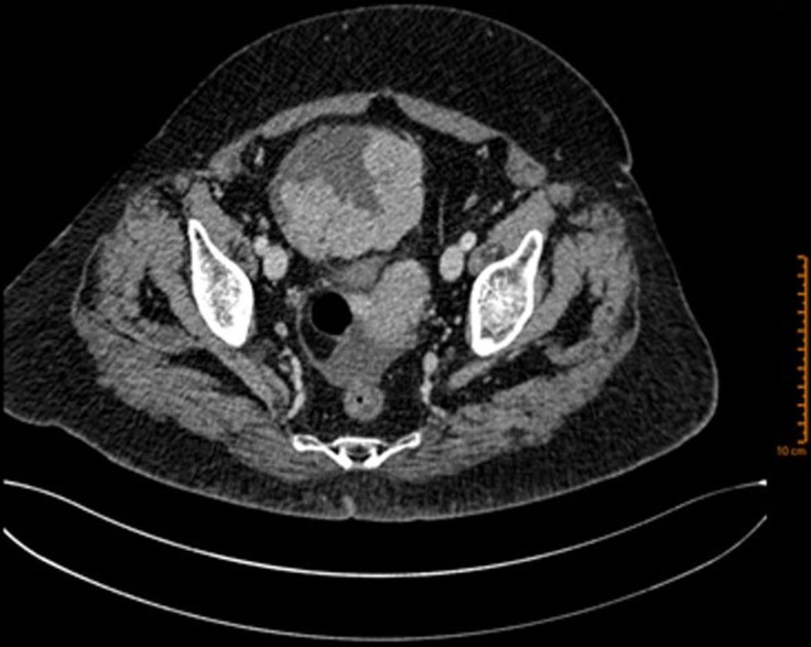
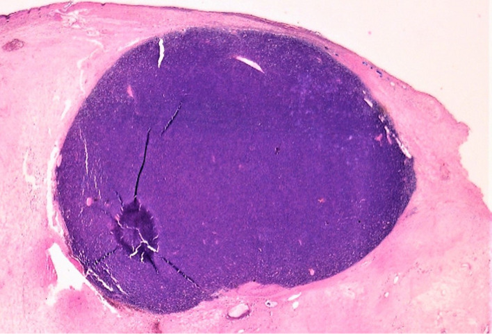
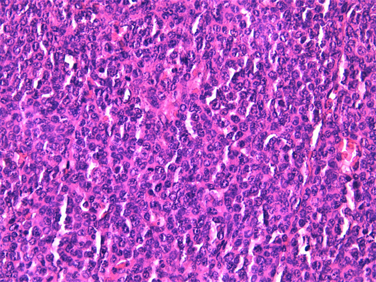
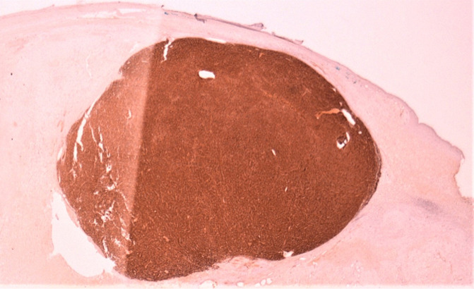
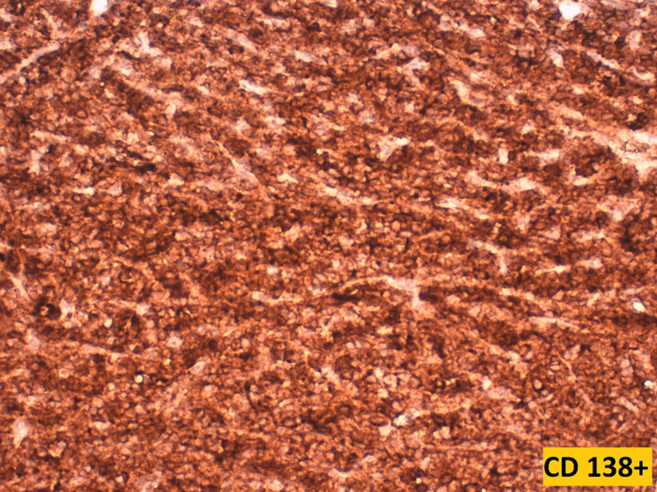
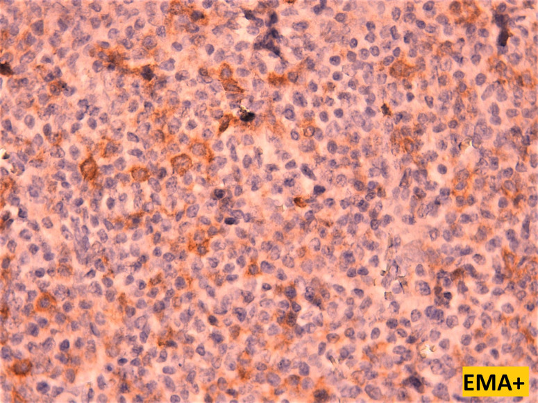
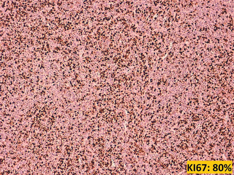
Plasmacytomas are a rare spectrum of plasma cell neoplasms that are single localized tumours, lacking the clinical features of plasma cell myeloma with no radiographical evidence of additional plasma cell tumours. Two clinical variants of plasmacytomas can be distinguished: solitary plasmacytoma of bone and extramedullary (or extraosseous) plasmacytoma. The latter is rare, representing 1% of all plasma cell neoplasms, occurring most frequently in the upper airways. Ovarian localization is exceptional, with only a few cases being reported in the literature. We herein report a case of an ovarian extramedullary plasmacytoma occurring in a 56-year-old woman who consulted for abdominal pain and abdominal mass, while highlighting the main histological and immunohistochemical features of this rare malignancy, along with a thorough review of literature gathering all cases of ovarian plasmacytomas reported to date.
Keywords: Extramedullary plasmacytoma; case report; ovary; plasma cell neoplasm.
Copyright: Anass Haloui et al.
Conflict of interest statement
The authors declare no competing interests.
Figures
References
-
- Ben Salah H, Hdiji S, Makni S, Ghorbel AM, Boudawara T, Elloumi M, et al. [Extramedullary plasmocytomas] Cancer Radiother. 2012 Jul-Aug;16(4):282–7. - PubMed
-
- Andze G, Pagbe JJ, Tchokoteu PF, Mbakop A, Yomi J, Juimo AG, et al. Le plasmocytome solitaire extraosseux ovarien: à propos d'un cas exceptionnel chez une enfant de 12 ans. J Chir (Paris) 1993 Mar;130(3):137–40. - PubMed
-
- Talerman A, Path FR. Nonspecific tumors of the ovary, including mesenchymal tumors and malignant lymphoma. Blaustein´s Pathology of the Female Genital Tract. In: RJ Kurman., editor. 3rd edn. New York: Springer-Verlag; 1987. pp. 722–41.
-
- Voegt H. Extramedullary plasmacytoma. Virchows Arch Pathol Anat. 1938;302:497–508.
Publication types
MeSH terms
LinkOut - more resources
Full Text Sources
Medical
