. 2023 May 12:29:439-449.
doi: 10.1016/j.omtm.2023.05.010. eCollection 2023 Jun 8.

Enzyme replacement with transferrin receptor-targeted α-L-iduronidase rescues brain pathology in mucopolysaccharidosis I mice

Affiliations

Enzyme replacement with transferrin receptor-targeted α-L-iduronidase rescues brain pathology in mucopolysaccharidosis I mice

Sachiho Kida et al. Mol Ther Methods Clin Dev. .

Abstract

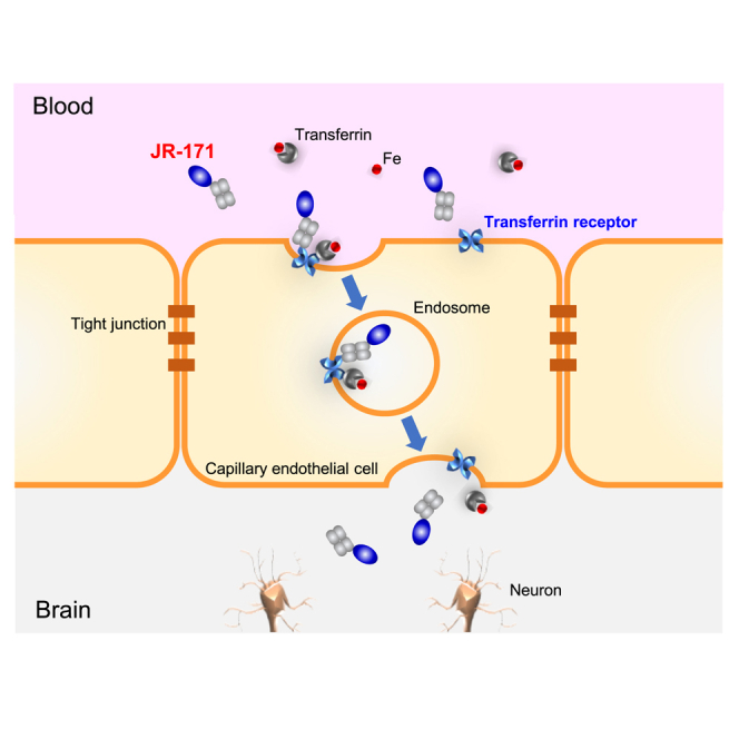
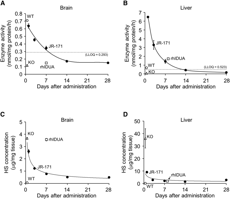
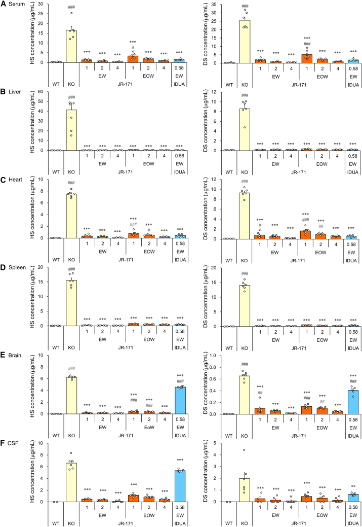
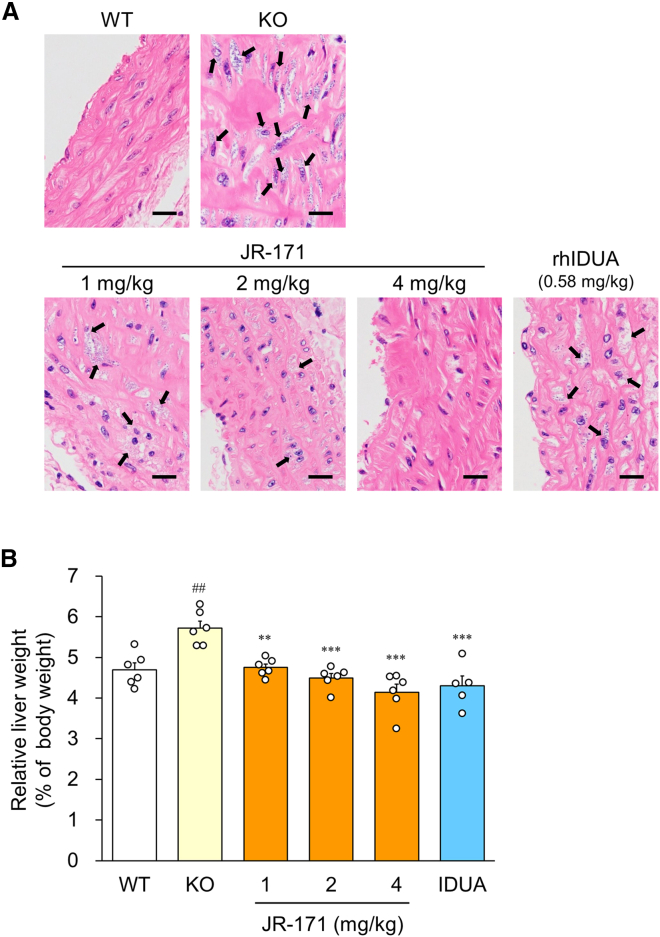
Mucopolysaccharidosis I (MPS I), a lysosomal storage disease caused by dysfunction of α-L-iduronidase (IDUA), is characterized by the deposition of dermatan sulfate (DS) and heparan sulfate (HS) throughout the body, which causes several somatic and central nervous symptoms. Although enzyme-replacement therapy (ERT) is currently available to treat MPS I, it does not alleviate central nervous disorders, as it cannot penetrate the blood-brain barrier. Here we evaluate the brain delivery, efficacy, and safety of JR-171, a fusion protein comprising humanized anti-human transferrin receptor antibody Fab and IDUA, using monkeys and MPS I mice. Intravenously administered JR-171 was distributed in major organs, including the brain, and reduced DS and HS concentrations in the central nervous system and peripheral tissues. JR-171 exerted similar effects on peripheral disorders similar to conventional ERT and further reversed brain pathology in MPS I mice. We found that JR-171 improved spatial learning ability, which was seen to deteriorate in the vehicle-treated mice. Further, no safety concerns were noted in repeat-dose toxicity studies in monkeys. This study provides nonclinical evidence that JR-171 might potentially prevent and even improve disease conditions in patients with neuronopathic MPS I without serious safety concerns.

Keywords: blood-brain barrier; enzyme-replacement therapy; heparan sulfate; lysosomal storage disease; mucopolysaccharidosis I.


Conflict of interest statement

S.K., Y.K., E.Y., A.Y., H.M., A. Imakiire, N.T., S.T., A.M., J.I., A. Inoue, R.Y., K.M., T.H., K.T., and H.S. are employees and/or stockholders of JCR Pharmaceuticals Co., Ltd.

Figures

None
Graphical abstract
Figure 1
Figure 1
Molecular structure and receptor-targeting property of JR-171 (A) Depicted molecular structure of JR-171. JR-171 is a recombinant fusion protein consisting of two subunits: a VH-CH1 heavy chain of a humanized anti-human transferrin receptor antibody C-terminally fused to IDUA via a glycine-serine linker and the light chain of the same antibody. Its molecular weight is calculated to be 119,204 Da based on its amino acid composition (C5337H8209N1479O1578S27). The amino acid sequence of the IDUA moiety is shown in the box. N, sugar chain binding asparagine; C-C, disulfide bond. (B) Receptor-mediated cellular uptake of JR-171 into human fibroblasts. JR-171 (160 nmol/L) was incubated with MRC-5 cells with or without M6P (10 mM) and/or the humanized anti-hTfR monoclonal antibody (400 μg/mL). Intracellular concentration of JR-171 was calculated from enzyme activity using an artificial substrate 4-MU-α-Ido. Endogenous enzyme activity was negligible in this assay. The values are represented as the mean ± SEM (n = 3). ∗∗∗p < 0.001 (versus JR-171 alone, Dunnett’s test).
Figure 2
Figure 2
Pharmacokinetic and biodistribution of JR-171 and rhIDUA in MPS I mice (A) Drug concentration profiles of JR-171 (2 mg/kg) or rhIDUA (2 mg/kg) administered intravenously to hTfR-KI/Idua-KO (MPS I) mice. The values are represented as the mean ± SEM for each group (n = 4). (B) Brain distribution of JR-171 (4 mg/kg) or rhIDUA (4 mg/kg) administered to the mice using cerebellum sections stained with an anti-IDUA antibody. The bottom panel is a high-power view of the white dashed rectangle shown in the upper panel. Dotted signals are observed in Purkinje neurons. Scale bars, 50 μm.
Figure 3
Figure 3
Enzyme activity and HS concentration in the brain and liver after intravenous administration of JR-171 to MPS I mice MPS I mice were intravenously injected with JR-171 (1 mg/kg) or rhIDUA (0.58 mg/kg). The amount of injected IDUA enzyme is almost equivalent at molar basis. IDUA enzyme activity in the brain (A) and liver (B), and HS concentrations in the brain (C) and liver (D) are shown. Enzyme activity in KO mice was below the quantification limit. Data represent the means ± SEM (n = 3 at each time point). WT, wild-type mice; KO, untreated hTfR-KI/Idua-KO (MPS I) mice; LLOQ, the lower limit of quantification.
Figure 4
Figure 4
Substrate-lowering efficacy of JR-171 in MPS I mice Graphs showing HS (right) and DS (left) concentrations in the serum (A), liver (B), heart (C), spleen (D), brain (E), and (F) CSF. JR-171 or rhIDUA was administered intravenously to 11-week-old MPS I mice once every week (EW) or once every other week (EOW) for 12 weeks at doses indicated on the horizontal axis of each graph (in mg/kg). Tissues were collected 1 week (for EW groups) or 2 weeks (for EOW groups) after the last dose. Values are represented as the mean with SEM bar for each group (n = 5–6). Individual values are also plotted. ###p < 0.001; ##p < 0.01; #p < 0.05 (versus WT, unpaired t test), ∗∗∗p < 0.001; ∗∗p < 0.01 (versus KO, Dunnett’s test). WT, wild-type mice; KO, untreated hTfR-KI/Idua-KO (MPS I) mice.
Figure 5
Figure 5
Effect of JR-171 on peripheral pathology in MPS I mice JR-171 (1, 2, or 4 mg/kg) or rhIDUA (0.58 mg/kg) was administered intravenously to 11-week-old MPS I mice once every week for 12 weeks. (A) H&E-stained sections of the aortic wall of the heart. Vacuolation and swelling of interstitial and smooth muscle cells are observed. Scale bars, 20 μm. (B) Relative liver weight. Values are presented as the mean with SEM bar for each group (n = 5–6). Individual values are also plotted. ##p < 0.01 (versus WT, unpaired t test), ∗∗∗p < 0.001; ∗∗p < 0.01 (versus KO, Dunnett’s test). WT, wild-type mice; KO, hTfR-KI/Idua-KO (MPS I) mice.
Figure 6
Figure 6
Effects of JR-171 on brain pathology in MPS I mice JR-171 (1 mg/kg) or rhIDUA (0.58 mg/kg) was administered intravenously to 11-week-old MPS I mice once every week for 31 weeks. (A) Representative photomicrographs of cerebellum sections are shown. Brown signals indicate positive staining for Lamp1, GFAP, or Iba1. White arrows indicate vacuolated Iba1-positive microglial cells, and black arrows indicate vacuolation or swelling of Purkinje neurons (H&E). Scale bars, 20 μm. (B) Grading of histopathological changes. Each square indicates an individual animal.
Figure 7
Figure 7
Morris water maze performance in MPS I mice treated with JR-171 (A) Goal latency. (B–D) Probe trial performed on day 4. The time spent in the target quadrant (B), the number of platform crossings (C), and the moved distance (D) were measured. MPS I mice were chronically treated with JR-171 (1 mg/kg/wk) or rhIDUA (0.58 mg/kg) for 24 weeks. The time to reach the platform (goal latency) was measured thrice daily and the means were calculated daily for individual animals. Values are represented as the mean with SEM bar for each group (n = 7–9). ∗p < 0.05 (compared with the value on day 1, paired t test). The probe trial was performed on day 4 after removing the platform. Differences are not statistically significant between any two groups (one-way ANOVA). WT, wild-type mice; KO, untreated hTfR-KI/Idua-KO (MPS I) mice.

References

    1. Kubaski F., de Oliveira Poswar F., Michelin-Tirelli K., Matte U.D.S., Horovitz D.D., Barth A.L., Baldo G., Vairo F., Giugliani R. Mucopolysaccharidosis type I. Diagnostics. 2020;10:161. - PMC - PubMed
    1. Hampe C.S., Eisengart J.B., Lund T.C., Orchard P.J., Swietlicka M., Wesley J., McIvor R.S. Mucopolysaccharidosis type I: a review of the natural history and molecular pathology. Cells. 2020;9:1838. - PMC - PubMed
    1. Hampe C.S., Wesley J., Lund T.C., Orchard P.J., Polgreen L.E., Eisengart J.B., McLoon L.K., Cureoglu S., Schachern P., McIvor R.S. Mucopolysaccharidosis type I: current treatments, limitations, and prospects for improvement. Biomolecules. 2021;11:189. - PMC - PubMed
    1. Aldenhoven M., Wynn R.F., Orchard P.J., O'Meara A., Veys P., Fischer A., Valayannopoulos V., Neven B., Rovelli A., Prasad V.K., et al. Long-term outcome of Hurler syndrome patients after hematopoietic cell transplantation: an international multicenter study. Blood. 2015;125:2164–2172. - PubMed
    1. Aldenhoven M., Jones S.A., Bonney D., Borrill R.E., Coussons M., Mercer J., Bierings M.B., Versluys B., van Hasselt P.M., Wijburg F.A., et al. Hematopoietic cell transplantation for mucopolysaccharidosis patients is safe and effective: results after implementation of international guidelines. Biol. Blood Marrow Transplant. 2015;21:1106–1109. - PubMed

LinkOut - more resources