. 2023 May 10:5:100115.
doi: 10.1016/j.bioflm.2023.100115. eCollection 2023 Dec.

Silver-doped bioactive glass fibres as a potential treatment for wound-associated bacterial biofilms

Affiliations

Silver-doped bioactive glass fibres as a potential treatment for wound-associated bacterial biofilms

Sandeep Shirgill et al. Biofilm. .

Abstract

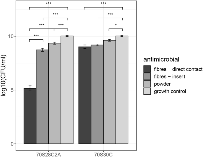
Chronic wounds are a drain on global health services and remain a major area of unmet clinical need. Chronic wounds are characterised by a stable and stubborn bacterial biofilm which hinders innate immune response and delays or prevents wound healing. Bioactive glass (BG) fibres offer a promising novel treatment for chronic wounds by targeting the wound-associated biofilm. In this study, the antimicrobial properties of silver-doped BG fibres were tested against Pseudomonas aeruginosa biofilms, which are commonly found in chronic wound infections. Results showed that BG fibres doped with silver resulted in a 5log10 reduction in biofilm formation whereas silver-free fibres only reduced formation by log10, therefore silver-doped fibres possess stronger antimicrobial effects. Moreover, there appeared to be a synergistic effect between the fibres and the silver as the application of the silver-doped fibres placed directly in contact with the forming biofilm resulted in a higher reduction in biofilm formation compared to treatments either: using the dissolution ions, using BG powder, or when the fibres were placed in an insert above the biofilm, inhibiting physical contact, instead. This suggests that the physical properties of the fibres, as well as silver, influence biofilm formation. Finally, results demonstrated that silver chloride, which is not antimicrobial, forms and the concentrations of antimicrobial silver species, namely silver ions and nanoparticles, reduce over time when fibres are soaked in cell culture media, which partially explains why the silver-doped dissolution ions contained lower antimicrobial activity compared to the fibres. As silver chloride is more likely to form with increased temperature and time, the antimicrobial activity of silver-containing dissolution ions is highly dependent on the length of ageing and storage conditions. Many studies investigate the antimicrobial and cytotoxic properties of biomaterials through their dissolution products. However, instability of antimicrobial silver species due to silver chloride formation and its effect on antimicrobial properties of silver-based biomaterials has not been reported before and could influence past and future dissolution-based assays as results showed that the antimicrobial activity of silver-based dissolution ions can vary greatly depending on post processing steps and can therefore produce misleading data.

Keywords: Antibacterial activity; Bioactive glass; Chronic wound biofilms; Pseudomonas aeruginosa; Silver.


Conflict of interest statement

The authors declare the following financial interests/personal relationships which may be considered as potential competing interests:Sandeep Shirgill reports financial support was provided by 10.13039/501100000268Biotechnology and Biological Sciences Research Council. Gowsihan Poologasundarampillai reports financial support was provided by 10.13039/501100000266Engineering and Physical Sciences Research Council.

Figures

Fig. 1
Fig. 1
Silicon species (Si), calcium (Ca) and silver (Ag) and changes in environmental pH during fibre dissolution up to 7 days. Both 70S30C and 70S28C2A fibres are soaked in RPMI 1640 in the ratio of 1.5 mg/ml. Error bars represent the average value ± 1 standard error of the mean.
Fig. 2
Fig. 2
Comparing the effect of placing fibres on inoculated membranes and using dissolution products on biofilm inhibition. Biofilm growth under these conditions was compared to a growth control of an untreated 24hr biofilm. Error bars represent the average value ± 1 standard error of the mean. Statistical significance was calculated using a 1-way ANOVA, where b × p-value 0.05, 0.005, 0.0005 is represented by, *, **, *** respectively (where b = 8 using the Bonferroni correction to correct for multiple comparisons).
Fig. 3
Fig. 3
XRD of the 70S30C and 70S28C2A fibres either soaked in RPMI 1640 for 7 days before analysis or unsoaked. The presence of diffraction peaks indicates crystalline structures in the fibres. Fig. 3a and b demonstrate 70S30C fibres before and after soaking respectively and Fig. 3c and d demonstrate 70S28C2A fibres before and after soaking respectively.
Fig. 4
Fig. 4
TEM images of the 70S30C and 70S28C2A fibres either soaked in RPMI 1640 for 7 days before imaging or unsoaked. Graphs show individual fibres and scale bars are 200 nm. Fig. 4a and b demonstrate 70S30C fibres before and after soaking respectively and Fig. 4c and d demonstrate 70S28C2A fibres before and after soaking respectively. Arrows on Fig. 4b and d indicate the locations from where SAED patterns were taken.
Fig. 5
Fig. 5
SAED patterns and measured d-spacing of fibres either soaked or not soaked in RPMI 1640 for 7 days. Fig. 5a is the corresponding diffraction pattern for soaked 70S30C fibres in Fig. 4b, where the diffraction pattern corresponds to HCA. Fig. 5b is the diffraction pattern for 70S28C2A fibres before soaking, where the d-spacing of rings corresponds to silver oxide. Finally Fig. 5c is the diffraction pattern for soaked 70S28C2A fibres seen in Fig. 4d, which identifies the crystal structure as silver chloride. Scale bars are of length 5 1/nm.
Fig. 6
Fig. 6
The change in silver ions and silver nanoparticle species concentration over time whilst dissolution products of 70S28C2A fibres are incubated at 37 °C. Error bars represent the average value ± 1 standard error of the mean.
Fig. 7
Fig. 7
Growth of planktonic PAO1-N, when exposed to dissolution products of standard fibres (Fig. 7b) or fibres doped with silver (Fig. 7a). Graphs compare the effect of age on dissolution products' efficacy as well as the effect of the temperature of storage conditions. Error bars represent the average value ± 1 standard error of the mean. Statistical significance was calculated using a 1-way ANOVA, where b × p-value 0.05, 0.005, 0.0005 is represented by, *, **, *** respectively (where b = 11 using the Bonferroni correction to correct for multiple comparisons).
Fig. 8
Fig. 8
The effect of BG on PA01 biofilm inhibition. Both 70S30C and 70S28C2A compositions were tested either by placing fibres directly on top of the inoculated membrane, placing an insert suspended above the membrane, or using BG powder. Biofilm growth under these conditions was compared to a growth control of an untreated 24hr biofilm. Error bars represent the average value ± 1 standard error of the mean. Statistical significance was calculated using an 1-way ANOVA, where b × p-value 0.05, 0.005, 0.0005 is represented by, *, **, *** respectively (where b = 10 using the Bonferroni correction to correct for multiple comparisons).
Fig. 9
Fig. 9
SEM and TEM images illustrating the interaction between BG fibres and planktonic PA01-N. From the images, it can be shown that fibres can physically disrupt the bacteria and cause leakage of intracellular components.
Fig. S1a
Fig. S1a
SAED patterns for unsoaked 70S28C2A fibres taken at different areas demonstrating the presence of diffused rings near the main transmitted beam.
Fig. S1b
Fig. S1b
A calibration curve to determine the relationship between PA01-N growth measure in OD600 and in CFU/ml. From the graph, a line of best fit was used to determine that 1 OD = 8.789 x 10⁸CFU/ml with an R2 value of 0.9923.
Image 1

References

    1. Kerr Marion. Inpatient care for people with diabetes: the economic case for change. Tech. rep. London: NHS Diabetes. 2011 http://webarchive.nationalarchives.gov.uk/20130513172211/ http://www.diabetes.nhs.uk/document.php?o=3034
    1. Julian F. Guest, et al. Health economic burden that wounds impose on the National Health Service in the UK”. In: BMJ Open. 2015;5(12):2044–6055. issn. - PMC - PubMed
    1. James Garth A., et al. Biofilms in chronic wounds. In: Wound Repair Regen. 2008;16(1):37–44. - PubMed
    1. Bowler P.G., Bi Duerden, Armstrong David G. Wound microbiology and associated approaches to wound management. In: Clin Microbiol Rev. 2001;14(2):244–269. - PMC - PubMed
    1. Percival Steven L., et al. A review of the scientific evidence for biofilms in wounds. In: Wound Repair Regen. 2012;20(5):647–657. - PubMed