. 2023 Apr 11;9(5):915-926.
doi: 10.1021/acscentsci.2c01317. eCollection 2023 May 24.

Rational Chemical Design of Molecular Glue Degraders

Affiliations

Rational Chemical Design of Molecular Glue Degraders

Ethan S Toriki et al. ACS Cent Sci. .

Erratum in

  • Correction to "Rational Chemical Design of Molecular Glue Degraders".
    Toriki ES, Papatzimas JW, Nishikawa K, Dovala D, Frank AO, Hesse MJ, Dankova D, Song JG, Bruce-Smythe M, Struble H, Garcia FJ, Brittain SM, Kile AC, McGregor LM, McKenna JM, Tallarico JA, Schirle M, Nomura DK. Toriki ES, et al. ACS Cent Sci. 2023 Jul 21;9(8):1702. doi: 10.1021/acscentsci.3c00844. eCollection 2023 Aug 23. ACS Cent Sci. 2023. PMID: 37637749 Free PMC article.

Abstract

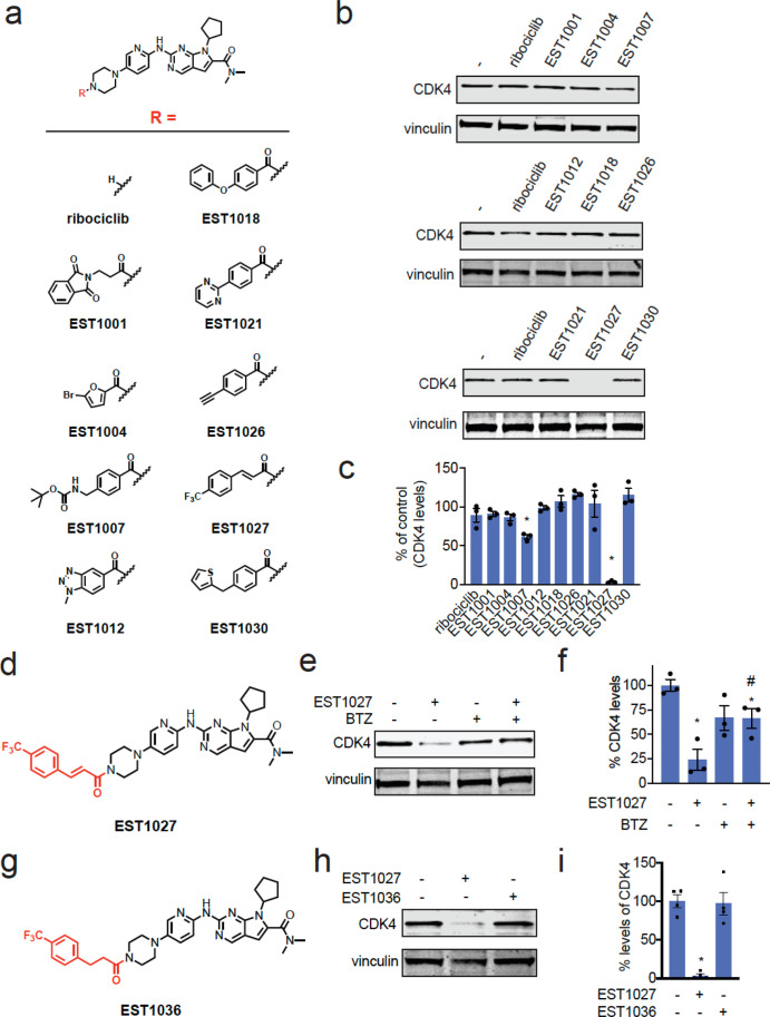
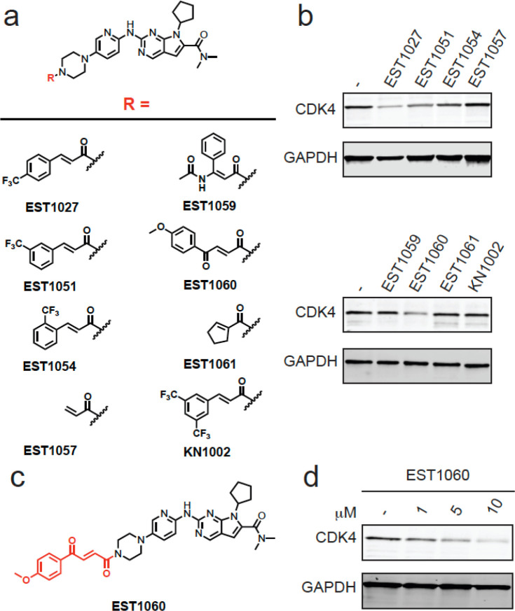
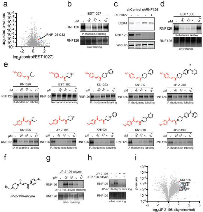
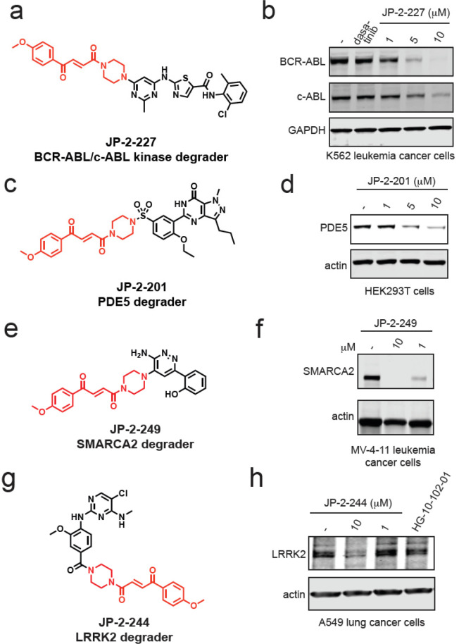
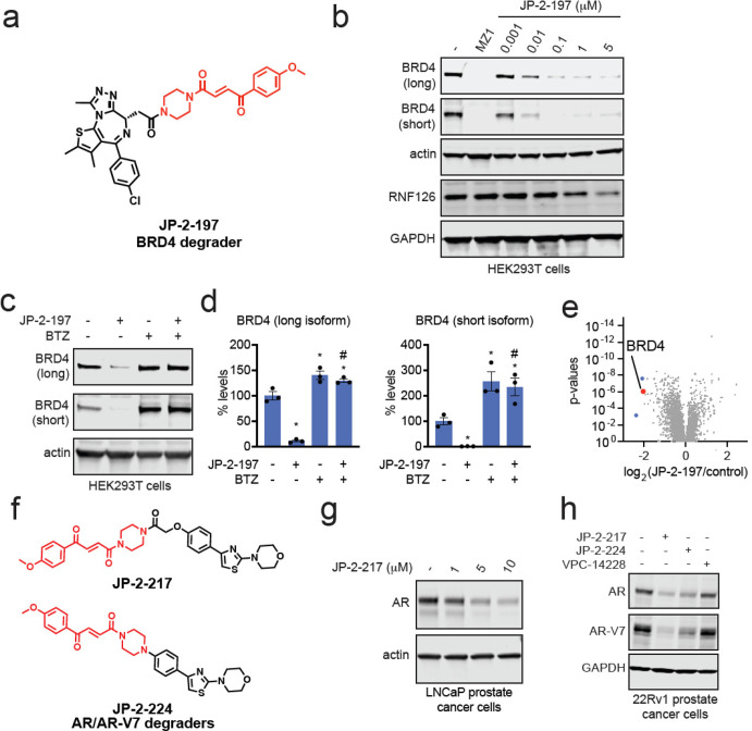
Targeted protein degradation with molecular glue degraders has arisen as a powerful therapeutic modality for eliminating classically undruggable disease-causing proteins through proteasome-mediated degradation. However, we currently lack rational chemical design principles for converting protein-targeting ligands into molecular glue degraders. To overcome this challenge, we sought to identify a transposable chemical handle that would convert protein-targeting ligands into molecular degraders of their corresponding targets. Using the CDK4/6 inhibitor ribociclib as a prototype, we identified a covalent handle that, when appended to the exit vector of ribociclib, induced the proteasome-mediated degradation of CDK4 in cancer cells. Further modification of our initial covalent scaffold led to an improved CDK4 degrader with the development of a but-2-ene-1,4-dione ("fumarate") handle that showed improved interactions with RNF126. Subsequent chemoproteomic profiling revealed interactions of the CDK4 degrader and the optimized fumarate handle with RNF126 as well as additional RING-family E3 ligases. We then transplanted this covalent handle onto a diverse set of protein-targeting ligands to induce the degradation of BRD4, BCR-ABL and c-ABL, PDE5, AR and AR-V7, BTK, LRRK2, HDAC1/3, and SMARCA2/4. Our study undercovers a design strategy for converting protein-targeting ligands into covalent molecular glue degraders.


Conflict of interest statement

The authors declare the following competing financial interest(s): JAT, JMK, MS, DD, ACK, HS, AOF, MBS, HS, SMB, FJG, MJH, and LM are employees of Novartis Institutes for BioMedical Research. JP was a University of California, Berkeley employee during this study, but is now a Novartis Institutes for BioMedical Research employee. This study was funded by the Novartis Institutes for BioMedical Research and the Novartis-Berkeley Translational Chemical Biology Institute. DKN is a co-founder, shareholder, and scientific advisory board member for Frontier Medicines and Vicinitas Therapeutics. DKN is a member of the board of directors for Vicinitas Therapeutics. DKN is also on the scientific advisory board of The Mark Foundation for Cancer Research, MD Anderson Cancer Center, Photys Therapeutics, Apertor Pharmaceuticals, Oerth Bio, and Chordia Therapeutics. DKN is also an Investment Advisory Board Member for Droia Ventures and a16z.

Figures

Figure 1
Figure 1
Synthesis and testing of analogs with chemical handles appended to the exit vector of the CDK4/6 inhibitor ribociclib. (a) Structures of ribociclib analogs wherein various chemical handles were appended onto the solvent-exposed end of ribociclib. (b) Testing ribociclib analogs in C33A cervical cancer cells to identify compounds that reduce CDK4 levels. C33A cells were treated with DMSO vehicle or compounds (3 μM) for 24 h. CDK4 and loading control vinculin levels were assessed by Western blotting. (c) Quantification of the data shown in (b). (d) Full structure of hit compound EST1027 that showed >50% loss of CDK4 in (b,c) with the appended chemical handle shown in red. (e) Proteasome-dependent degradation of CDK4 by EST1027. C33A cells were pretreated with DMSO vehicle or the proteasome inhibitor bortezomib (BTZ) (10 μM) 1 h prior to treatment of cells with DMSO vehicle or EST1027 (5 μM), and CDK4 and loading control vinculin levels were assessed by Western blotting. (f) Quantification of the experiment described in (e). (g) Structure of EST1036, a nonreactive derivative of EST1027. (h) EST1036 does not degrade CDK4. C33A cells were treated with DMSO vehicle or compounds (5 μM) for 24 h, and CDK4 and loading control vinculin levels were assessed by Western blotting. (i) Quantification of experiment in (h). Blots shown in (b,e,h) are representative of n = 3 biologically independent replicates/group. Bar graphs in (c,f,i) show individual replicate values and average ± sem. Statistical significance is calculated as *p < 0.05 compared to DMSO vehicle in (c,f,i) and #p < 0.05 compared to the EST1027-treated group in (f).
Figure 2
Figure 2
Structure–activity relationship of CDK4 degrader. (a) Structures of EST1027 analogs assessing structure–activity relationships. (b) Testing EST1027 analogs in C33A cervical cancer cells to identify compounds that reduce CDK4 levels. C33A cells were treated with DMSO vehicle or compounds (5 μM) for 24 h. CDK4 and loading control GAPDH levels were assessed by Western blotting. (c) Full structure of hit compound EST1060. (d) Dose–response of EST1060 CDK4 degradation. C33A cells were treated with DMSO vehicle or EST1060 for 24 h. CDK4 and loading control GAPDH levels were assessed by Western blotting. Gels and blots in (b,d) are representative images from n = 3 biologically independent replicates/group.
Figure 3
Figure 3
Mapping proteome-wide interactions of the fumarate handle. (a) Cysteine chemoproteomic profiling of EST1027 in C33A cervical cancer cells using isoDTB-ABPP. C33A cells were treated with DMSO vehicle or EST1027 (20 μM) for 2 h. Resulting lysates were labeled with an alkyne-functionalized iodoacetamide probe (IA-alkyne) (200 μM) for 1 h, after which isotopic desthiobiotin tags were appended by copper-catalyzed azide–alkyne cycloaddition (CuAAC) and taken through the isoDTB-ABPP procedure. Shown in blue and red are probe-modified cysteines that showed control/EST1027 ratios >2 with p < 0.05 from n = 3 biologically independent replicates. Shown in red is RNF126 C32. (b) Gel-based ABPP of EST1027 against RNF126. Recombinant RNF126 was preincubated with DMSO vehicle or EST1027 for 30 min prior to labeling of RNF126 with IA-rhodamine (250 nM) for 1 h. Gels were visualized by in-gel fluorescence, and protein loading was assessed by silver staining. (c) RNF126 knockdown attenuates EST1027-mediated CDK4 degradation. RNF126 was stably knocked down in C33A cells using short hairpin oligonucleotides (shRNF126) compared to nontargeting shControl oligonucleotides. C33A shControl and shRNF126 cells were treated with DMSO vehicle or EST1027 (5 μM) for 24 h. CDK4, RNF126, and loading control vinculin levels were assessed by Western blotting. (d) Gel-based ABPP of EST1060 against RNF126 performed as described in (b). (e) Gel-based ABPP of covalent chemical handles against RNF126 performed as described in (b). (f) Structure of JP-2-196-alkyne probe. (g) JP-2-196-alkyne labeling of pure RNF126 protein. RNF126 was labeled with DMSO vehicle or JP-2-196-alkyne for 30 min. Probe-modified RNF126 was subjected to CuAAC with a rhodamine-functionalized azide handle and visualized by SDS/PAGE and in-gel fluorescence. (h) Competition of JP-2-196-alkyne labeling of RNF126 by JP-2-196. RNF126 pure protein was preincubated with JP-2-196 (50 μM) for 30 min at 37 °C prior to JP-2-196 labeling (50 μM) for 30 min at room temperature. Probe-modified RNF126 was subjected to CuAAC with a rhodamine-functionalized azide handle and visualized by SDS/PAGE and in-gel fluorescence. (i) JP-2-196-alkyne pulldown proteomics showing significant and moderately selective engagement of RNF126 and five additional E3 ubiquitin ligases LRSAM1, RNF40, MID2, RNF219, and RNF14. HEK293T cells were treated with DMSO vehicle or JP-2-196-alkyne (10 μM) for 6 h. Subsequent lysates were subjected to CuAAC with an azide-functionalized biotin handle, after which probe-modified proteins were avidin-enriched, eluted, and digested, and analyzed by TMT-based quantitative proteomics. Data shown are ratios of JP-2-196-alkyne vs DMSO-control-enriched proteins and p-values from n = 3 biologically independent replicates/group. Gels and blots from (b–e,g,h) are representative of n = 3 biologically independent replicates/group.
Figure 4
Figure 4
Transplanting a covalent chemical handle onto protein-targeting ligands that already possess piperazines at the exit vector. (a) Structure of JP-2-227 with the optimized covalent handle shown in red that was appended onto the BCR-ABL and c-ABL kinase inhibitor dasatinib. (b) JP-2-227 degrades BCR-ABL and c-ABL in K562 leukemia cancer cells. K562 cells were treated with DMSO vehicle or JP-2-227 for 24 h, and BCR-ABL, c-ABL, and loading control GAPDH levels were assessed by Western blotting. (c) Structure of JP-2-201 with the optimized covalent handle shown in red that was appended onto the PDE5 inhibitor sildenafil. (d) JP-2-201 degrades PDE5 in HEK293T cells. HEK293T cells were treated with DMSO vehicle or JP-2-201 for 24 h, and PDE5 and loading control actin levels were assessed by Western blotting. (e) Structure of SMARCA2 degrader JP-2-249 consisting of the optimized covalent handle incorporated into a previously reported protein-targeting ligand for SMARCA2. (f) MV-4-11 leukemia cancer cells were treated with DMSO vehicle or JP-2-249 for 24 h, and SMARCA2 and actin loading control levels were assessed by Western blotting. (g) Structure of LRRK2 degrader JP-2-244 consisting of the optimized covalent handle incorporated into a previously reported LRRK2 inhibitor. (h) A549 lung cancer cells were treated with DMSO vehicle or JP-2-244 for 24 h, and LRRK2 and actin loading control levels were assessed by Western blotting. Blots in (b,d,f,h) are representative of n = 3 biologically independent replicates/group.
Figure 5
Figure 5
Transplanting a covalent chemical handle onto BET family inhibitor JQ1 and AR/AR-V7 targeting ligand to degrade BRD4 and AR/AR-V7. (a) Structure of JP-2-197 with the optimized covalent handle shown in red that was appended onto the BET family bromodomain inhibitor JQ1. (b) JP-2-197 degrades BRD4 in HEK293T cells. HEK293T cells were treated with DMSO vehicle, positive control BRD4 PROTAC MZ1 (1 μM), or JP-2-197 for 24 h. BRD4 long and short isoforms, RNF126, and loading controls actin and GAPDH levels were assessed by Western blotting. (c) Proteasome-dependent degradation of BRD4 by JP-2-197. HEK293T cells were pretreated with DMSO vehicle or the proteasome inhibitor BTZ (10 μM) for 1 h prior to treatment of cells with DMSO vehicle or JP-2-197 (1 μM), and BRD4 and loading control actin levels were assessed by Western blotting. (d) Quantification of the experiment described in (c). (e) TMT-based quantitative proteomic profiling of JP-2-197 in HEK293T cells. HEK293T cells were treated with DMSO vehicle or JP-2-197 (1 μM) for 24 h. Data are from n = 2 biological replicates per group. (f) Structures of two AR-V7 degraders consisting of the fumarate handle linked to an AR DNA-binding domain ligand VPC-14228–JP-2-217 and JP-2-224. (g) LNCaP prostate cancer cells were treated with JP-2-217 for 24 h, and AR and loading control actin levels were detected by Western blotting. (h) 22Rv1 prostate cancer cells were treated with DMSO vehicle, JP-2-217, JP-2-224, or VPC-14228 (10 μM) for 24 h, and AR, AR-V7, and loading control GAPDH levels were assessed by Western blotting. Blots and gels shown in (b,c,g,h) are representative images from n = 3 biologically independent replicates. Bar graphs in (d) show individual replicate values and average ± sem. Statistical significance is calculated as *p < 0.05 compared to DMSO vehicle and #p < 0.05 compared to cells treated with JP-2-197 alone.

References

    1. Bond M. J.; Crews C. M. Proteolysis targeting chimeras (PROTACs) come of age: entering the third decade of targeted protein degradation. RSC Chem. Biol. 2021, 2, 725–742. 10.1039/D1CB00011J. - DOI - PMC - PubMed
    1. Hughes S. J.; Ciulli A. Molecular recognition of ternary complexes: a new dimension in the structure-guided design of chemical degraders. Essays Biochem. 2017, 61, 505–516. 10.1042/EBC20170041. - DOI - PMC - PubMed
    1. Schreiber S. L. The Rise of Molecular Glues. Cell 2021, 184, 3–9. 10.1016/j.cell.2020.12.020. - DOI - PubMed
    1. Burslem G. M.; Crews C. M. Small-Molecule Modulation of Protein Homeostasis. Chem. Rev. 2017, 117, 11269–11301. 10.1021/acs.chemrev.7b00077. - DOI - PubMed
    1. Chamberlain P. P.; Hamann L. G. Development of targeted protein degradation therapeutics. Nat. Chem. Biol. 2019, 15, 937–944. 10.1038/s41589-019-0362-y. - DOI - PubMed