. 2023 Mar 16;9(5):870-882.
doi: 10.1021/acscentsci.3c00052. eCollection 2023 May 24.

Dihydroxy-Metabolites of Dihomo-γ-linolenic Acid Drive Ferroptosis-Mediated Neurodegeneration

Affiliations

Dihydroxy-Metabolites of Dihomo-γ-linolenic Acid Drive Ferroptosis-Mediated Neurodegeneration

Morteza Sarparast et al. ACS Cent Sci. .

Abstract

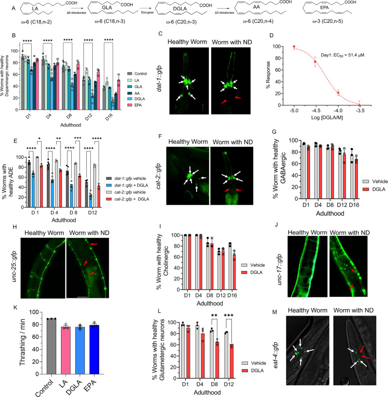
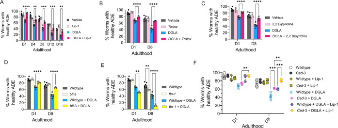
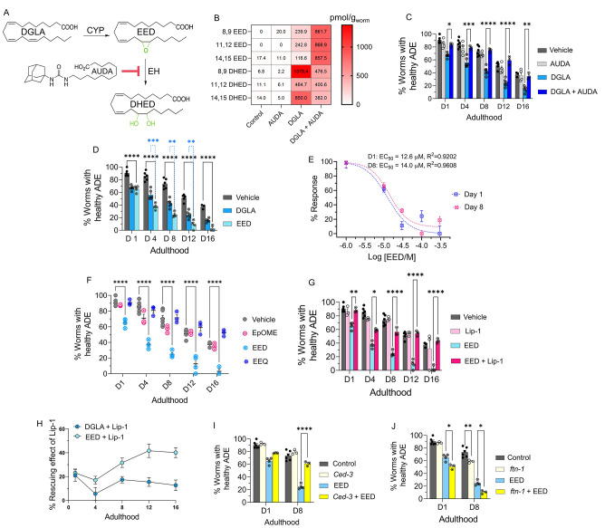
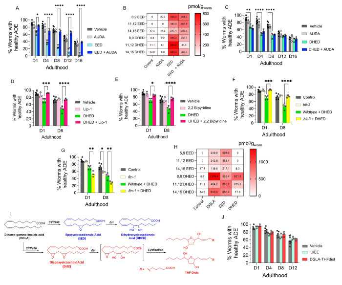
Even after decades of research, the mechanism of neurodegeneration remains understudied, hindering the discovery of effective treatments for neurodegenerative diseases. Recent reports suggest that ferroptosis could be a novel therapeutic target for neurodegenerative diseases. While polyunsaturated fatty acid (PUFA) plays an important role in neurodegeneration and ferroptosis, how PUFAs may trigger these processes remains largely unknown. PUFA metabolites from cytochrome P450 and epoxide hydrolase metabolic pathways may modulate neurodegeneration. Here, we test the hypothesis that specific PUFAs regulate neurodegeneration through the action of their downstream metabolites by affecting ferroptosis. We find that the PUFA dihomo-γ-linolenic acid (DGLA) specifically induces ferroptosis-mediated neurodegeneration in dopaminergic neurons. Using synthetic chemical probes, targeted metabolomics, and genetic mutants, we show that DGLA triggers neurodegeneration upon conversion to dihydroxyeicosadienoic acid through the action of CYP-EH (CYP, cytochrome P450; EH, epoxide hydrolase), representing a new class of lipid metabolites that induce neurodegeneration via ferroptosis.


Conflict of interest statement

The authors declare no competing financial interest.

Figures

Figure 1
Figure 1
DGLA, but not other ω-3 and ω-6 PUFAs, induces degeneration, specifically in dopaminergic neurons. (A) Structure of different ω-6 and ω-3 PUFAs examined in this study. (B) Percentage (%) of worms with healthy dopaminergic neurons for Pdat-1::gfp with and without supplementation with 100 μM of different ω-6 and ω-3 PUFAs. (C) Fluorescent images of Pdat-1::gfp worms with healthy and degenerated dopaminergic neurons (white arrows represent healthy neurons, and red arrows show degenerated/disappeared neurons). (D) Dose response curve: the effect of different DGLA concentrations on degeneration of ADE neurons on day 1 adulthood. (E) Comparison of the ADE neuron degeneration in Pdat-1::gfp and Pcat-2::gfp supplemented with 100 μM DGLA. (F) Fluorescent images of Pcat-2::gfp worms with healthy and degenerated dopaminergic neurons (white arrows represent healthy neurons, and red arrows show degeneration/disappearance of neurons). (G) Percentage (%) of worms with healthy GABAergic neurons for Punc-25::gfp with and without supplementation with 100 μM DGLA. (H) Fluorescence images of Punc-25::gfp worm with healthy and degenerated GABAergic neurons (red arrows show different signs of neurodegeneration including ventral cord break, commissure break, and branches). (I) Percentage (%) of worms with healthy cholinergic neurons for Punc-17::gfp with and without supplementation with 100 μM DGLA. (J) Fluorescence images of Punc-17::gfp worms with healthy and degenerated cholinergic neurons (red arrows show different signs of neurodegeneration including ventral cord break, commissure break, and branches). (K) Thrashing on day 8 adulthood of wild-type raised on 100 μM LA, DGLA, and EPA. (L) Percentage (%) of worms with healthy glutamatergic neurons with Peat-4::gfp with and without supplementation with 100 μM DGLA. (M) Fluorescent images of Peat-4::gfp worms with healthy and degenerated glutamatergic neurons (white arrows represent healthy neurons, and red arrows show degenerated/disappeared neurons). All supplementations were done at the L4 stage. For all experiments, N = 3, and about 20 worms were tested on each trial. Two-way analysis of variance (ANOVA) and Tukey’s multiple comparison test for panels B and D; t test for K: *P ≤ 0.05, **P ≤ 0.01, ***P ≤ 0.001, ****P < 0.0001, nonsignificant is not shown.
Figure 2
Figure 2
DGLA induces neurodegeneration in dopaminergic neurons via ferroptosis. (A) Percentage (%) of worms with healthy ADE neurons of worms exposed to 100 μM DGLA ± 250 μM liproxstatin-1. (B) Percentage (%) of worms with healthy ADE neurons in wild-type C. elegans treated with 100 μM DGLA ± 500 μM Trolox (vitamin E). (C) Percentage (%) of worms with healthy ADE neurons for Pdat-1::gfp worms treated with 100 μM DGLA ± 100 μM 2,2′-bipyridine. (D) Percentage (%) of worms with healthy ADE neurons in Pdat-1::gfp and Pdat-1::gfp;bli-3 worms treated with 100 μM DGLA. (E) Percentage (%) of worms with healthy ADE neurons for Pdat-1::gfp and Pdat-1::gfp;ftn-1 worms treated with 100 μM DGLA. (F) Percentage (%) of worms with healthy ADE neurons with Pdat-1::gfp and Pdat-1::gfp;ced-3 worms treated with 100 μM DGLA ± 250 μM liproxstatin-1. All supplementations were done at the L4 stage. Two-way analysis of variance (ANOVA), Tukey’s multiple comparison test. *P ≤ 0.05, **P ≤ 0.01, ***P ≤ 0.001, ****P < 0.0001; NS, not significant. DGLA, Dihomo-γ-linolenic acid; LA, linoleic acid; EPA, eicosapentaenoic acid; Lip-1, liproxstatin-1.
Figure 3
Figure 3
EED, epoxy metabolites downstream of DGLA, induce neurodegeneration by ferroptosis. (A) DGLA is metabolized to EED and DHED through the CYP and epoxide hydrolase enzymes, respectively, and AUDA inhibits epoxide hydrolase. (B) Oxylipin profile representing the pmol/g of EED and DHED regioisomers in worms treated with 100 μM DGLA ± 100 μM AUDA compared to control. (C) Percentage (%) of worms with healthy ADE neurons for Pdat-1::gfp treated with 100 μM DGLA ± 100 μM AUDA. (D) Percentage (%) of worms with healthy ADE neurons for Pdat-1::gfp worms treated with 100 μM DGLA and 100 μM EED. (E) Dose response curve: effect of different concentrations of EED on degeneration of ADE neurons on day 1 and day 8 adulthood. (F) Percentage (%) of worms with healthy ADE neurons in Pdat-1::gfp worms treated with 100 μM of different Ep-PUFAs, EpOME, and EEQ. (G) Percentage (%) of worms with healthy ADE neurons of worms treated with 100 μM DGLA ± 100 μM liproxstatin-1. (H) Comparison of the effect of 250 μM liproxstatin-1 on Pdat-1::gfp worms treated with 100 μM DGLA compared to 100 μM EED. (I) Percentage (%) of worms with healthy ADE neurons with Pdat-1::gfp and Pdat-1::gfp;ced-3 worms treated with 100 μM. (J) Percentage (%) of worms with healthy ADE neurons for Pdat-1::gfp and Pdat-1::gfp;ftn-1 worms treated with 100 μM DGLA; all supplementations were done at the L4 stage. Two-way analysis of variance (ANOVA), Tukey’s multiple comparison test. *P ≤ 0.05, **P ≤ 0.01, ***P ≤ 0.001, ****P < 0.0001; without *, not significant. DGLA, Dihomo-γ-linolenic acid; EED, epoxyeicosadienoic acids; DHED, dihydroxyeicosadienoic acids; CYP, cytochrome P450; EH, epoxide hydrolase; AUDA, 12-(1-adamantane-1-yl-ureido-) dodecanoic acid; Lip-1, liproxstatin-1; EpOME, epoxyoctadecenoic acids; EEQ, epoxyeicosatetraenoic acid.
Figure 4
Figure 4
DHED, dihydroxy fatty acid downstream of DGLA/EED, is key candidates for neurodegeneration induced by DGLA in dopaminergic neurons. (A) Percentage (%) of worms with healthy ADE neurons in Pdat-1::gfp worms treated with 100 μM EED ± 100 μM AUDA. (B) Oxylipin profile representing pmol/g of EED and DHED regioisomers in worms treated with 100 μM EED ± 100 μM AUDA compared to control. (C) Percentage (%) of worms with healthy ADE neurons for Pdat-1::gfp treated with 100 μM DHED ± 100 μM AUDA. (D) Percentage (%) of worms with healthy ADE neurons of worms exposed to 100 μM DHED ± 250 μM liproxstatin-1. (E) Percentage (%) of worms with healthy ADE neurons for Pdat-1::gfp worms treated with 100 μM DHED ± 100 μM 2,2′-bipyridine. (F) Percentage (%) of worms with healthy ADE neurons for Pdat-1::gfp and Pdat-1::gfp;ftn-1 worms treated with 100 μM DHED. (G) Percentage (%) of worms with healthy ADE neurons in Pdat-1::gfp and Pdat-1::gfp;bli-3 worms treated with 100 μM DHED. (H) Oxylipin profile representing the pmol/g of EED and DHED regioisomers in worms treated with 100 μM DGLA, EED, and DHED compared to control. (I) Two possible metabolisms of DGLA through the CYP/EH pathways; the alternative metabolism is that CYP can do two consecutive oxidations (or under oxidative stress) to yield diepoxies EED, after which EH will open one epoxide which under physiological conditions can cyclize to THF diols. (J) Percentage (%) of worms with healthy ADE neurons for Pdat-1::gfp treated with 100 μM DiEE and 100 μM DGLA-THF diol. All supplementations were done at the L4 stage. Two-way analysis of variance (ANOVA), Tukey’s multiple comparison test. *P ≤ 0.05, **P ≤ 0.01, ***P ≤ 0.001, ****P < 0.0001; without *, not significant. DGLA, Dihomo-γ-linolenic acid; EED, epoxyeicosadienoic acid; DHED, dihydroxyeicosadienoic acid; CYP, cytochrome P450; EH, epoxide hydrolase; AUDA, 12-(1-adamantane-1-yl-ureido-) dodecanoic acid; DiEE, diepoxyeicosadienoic acid.

Update of

References

    1. United Nations Department of Economic and Social Affairs . References. In World Population Ageing 2019; UN, 2020; pp 37–38.10.18356/7f55a58e-en. - DOI
    1. Hou Y.; Dan X.; Babbar M.; Wei Y.; Hasselbalch S. G.; Croteau D. L.; Bohr V. A. Ageing as a Risk Factor for Neurodegenerative Disease. Nat. Rev. Neurol. 2019, 15 (10), 565–581. 10.1038/s41582-019-0244-7. - DOI - PubMed
    1. Gan L.; Cookson M. R.; Petrucelli L.; La Spada A. R. Converging Pathways in Neurodegeneration, from Genetics to Mechanisms. Nat. Neurosci. 2018, 21 (10), 1300–1309. 10.1038/s41593-018-0237-7. - DOI - PMC - PubMed
    1. Dixon S. J.; Lemberg K. M.; Lamprecht M. R.; Skouta R.; Zaitsev E. M.; Gleason C. E.; Patel D. N.; Bauer A. J.; Cantley A. M.; Yang W. S.; Morrison B.; Stockwell B. R. Ferroptosis: An Iron-Dependent Form of Nonapoptotic Cell Death. Cell 2012, 149 (5), 1060–1072. 10.1016/j.cell.2012.03.042. - DOI - PMC - PubMed
    1. Ren J.-X.; Sun X.; Yan X.-L.; Guo Z.-N.; Yang Y. Ferroptosis in Neurological Diseases. Front. Cell. Neurosci. 2020, 14, 218.10.3389/fncel.2020.00218. - DOI - PMC - PubMed