. 2023 Jun 6;120(23):e2300052120.
doi: 10.1073/pnas.2300052120. Epub 2023 May 30.

Glial dysregulation in the human brain in fragile X-associated tremor/ataxia syndrome

Affiliations

Glial dysregulation in the human brain in fragile X-associated tremor/ataxia syndrome

Caroline M Dias et al. Proc Natl Acad Sci U S A. .

Abstract

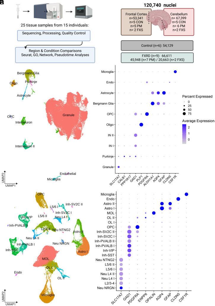
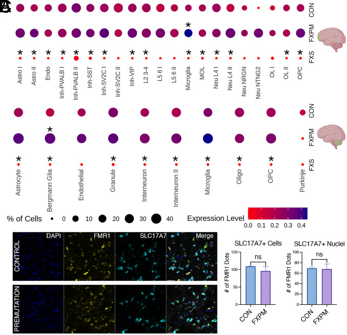
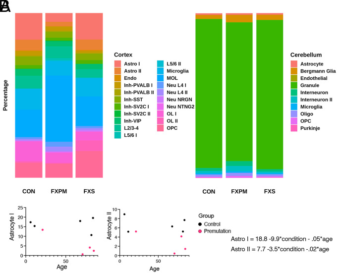
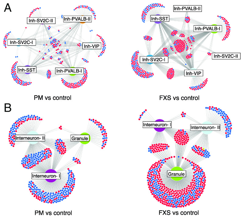
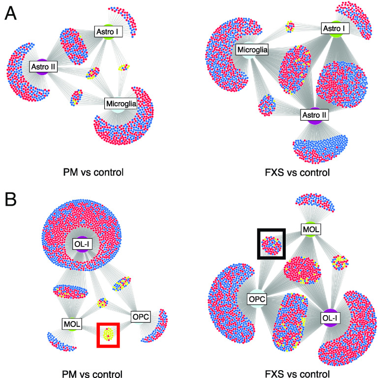
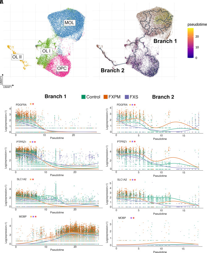
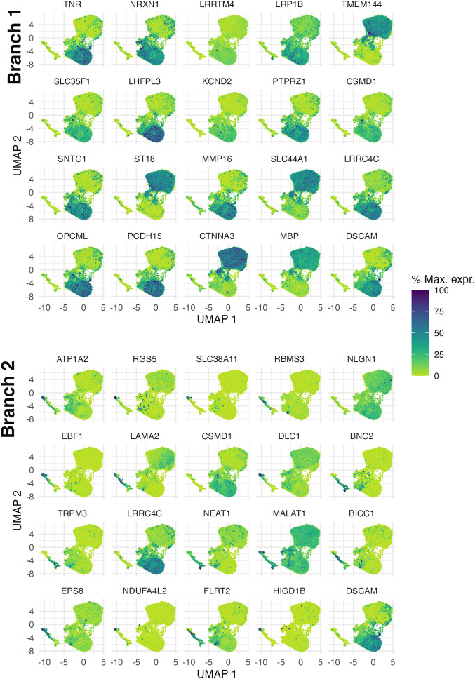
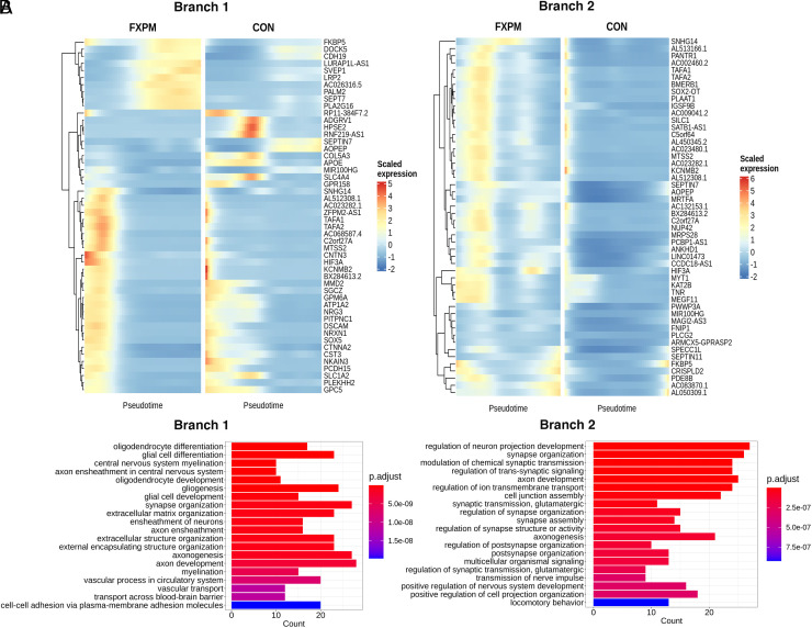
Short trinucleotide expansions at the FMR1 locus are associated with the late-onset condition fragile X-associated tremor/ataxia syndrome (FXTAS), which shows very different clinical and pathological features from fragile X syndrome (associated with longer expansions), with no clear molecular explanation for these marked differences. One prevailing theory posits that the shorter, premutation expansion uniquely causes extreme neurotoxic increases in FMR1 mRNA (i.e., four to eightfold increases), but evidence to support this hypothesis is largely derived from analysis of peripheral blood. We applied single-nucleus RNA sequencing to postmortem frontal cortex and cerebellum from 7 individuals with premutation and matched controls (n = 6) to assess cell type-specific molecular neuropathology. We found only modest upregulation (~1.3-fold) of FMR1 in some glial populations associated with premutation expansions. In premutation cases, we also identified decreased astrocyte proportions in the cortex. Differential expression and gene ontology analysis demonstrated altered neuroregulatory roles of glia. Using network analyses, we identified cell type-specific and region-specific patterns of FMR1 protein target gene dysregulation unique to premutation cases, with notable network dysregulation in the cortical oligodendrocyte lineage. We used pseudotime trajectory analysis to determine how oligodendrocyte development was altered and identified differences in early gene expression in oligodendrocyte trajectories in premutation cases specifically, implicating early cortical glial developmental perturbations. These findings challenge dogma regarding extremely elevated FMR1 increases in FXTAS and implicate glial dysregulation as a critical facet of premutation pathophysiology, representing potential unique therapeutic targets directly derived from the human condition.

Keywords: FMR1; FXTAS; glia; human brain; snRNA-seq.


Conflict of interest statement

The authors declare no competing interest.

Figures

Fig. 1.
Fig. 1.
Cell type–specific analysis of frontal cortex and cerebellum. (A) Sample preparation included dounce homogenization, sucrose centrifugation, fluorescent nuclear sorting, and nuclear encapsulation. (B) Summary of sample size and final filtered nuclei number per condition and region. (C) Cerebellar UMAP plot and dot plot of cell type–specific markers. (D) Frontal cortex UMAP plot and dot plot of cell type–specific markers in the frontal cortex. Abbreviations- FXRD: Fragile X-related disorders, Endo: endothelial, Astro: astrocyte, MOL: mature cortical oligodendrocyte, Oligo: mature cerebellar oligodendrocyte, OPC: oligodendrocyte progenitor, IN: interneuron, OL: oligodendrocyte lineage, Neu: excitatory neuron, Inh: inhibitory neuron, L: layer-specific excitatory neuron clusters.
Fig. 2.
Fig. 2.
Modest FMR1 changes in premutation postmortem brain. (A) snRNA-seq of frontal cortex and cerebellar changes in FMR1 expression in premutation cases and FXS cases vs control. Cluster abbreviations as in Fig. 1. In premutation cases, only cerebellar Bergmann glia and cortical microglia demonstrated significant increases in FMR1 mRNA expression. There was widespread FMR1 reduction in FXS cases despite the smaller sample size. (Asterisk above the dot indicates padj < 0.05 for condition vs control comparison.) Expression level indicates average of scaled log-normalized expression. (B) Representative image from fluorescent in-situ hybridization demonstrating comparable FMR1 expression in excitatory neurons (SLC17A7 marker) in premutation vs control in the frontal cortex. (Scale bar, 30 μm). (C) No significant difference in FMR1 mRNA was seen in SLC17A7+ nuclei, or, using an extended boundary, SLC17A7+ cells (two-tailed t test, P = 0.91 (nuclei), P = 0.46 (cells)). n = 2 control, n = 3 premutation.
Fig. 3.
Fig. 3.
Change in cellular abundance in postmortem brain. (A) Average percentage of nuclear composition in the frontal cortex and cerebellum. (B) For premutation and control samples, linear regression analysis was conducted to determine the effect of condition and age on cellular abundance, using the equation cluster % = β0 + β1*condition + β2*age. There was a significant effect of premutation condition on both cortical astrocyte groups (β1 P < 0.05), but not age (β2 P > 0.05). 89+ aged individual is omitted from graph but was included in regression analysis.
Fig. 4.
Fig. 4.
Network analysis of neuronal FMRP target dysregulation. Frontal cortex inhibitory neurons (A) demonstrate a hub of common derepressed FMRP target genes in FXS (Right), which is not observed in premutation cases (Left). (B) Cerebellar neurons in FXS also demonstrate a shared derepressed hub, with an opposite pattern in premutation cases. Different cell types demonstrate disproportionate effects of FMRP dysregulation depending on mutation status. Cluster abbreviations as in Fig. 1. Red, upregulation; blue, downregulation; yellow, opposite regulation in different cell types.
Fig. 5.
Fig. 5.
Network analysis of cortical glial FMRP target dysregulation. (A) Frontal cortex astrocytes and microglia demonstrate cell type–specific effects of FMRP network dysregulation in premutation cases (Left) and FXS (Right) (B) Oligodendrocyte lineage in premutation cases demonstrate uniquely divergent regulation of FMRP targets between OPCs and MOLs (red box); this pattern is not seen in FXS (black box). Cluster abbreviations as in Fig. 1. Red, upregulation; blue, downregulation; yellow, opposite regulation in different cell types.
Fig. 6.
Fig. 6.
Pseudotime analysis of cortical oligodendrocytes. (A) Reclustering of major oligodendrocyte clusters identifies two major trajectory branches identified in the frontal cortex. (A) Expression of oligodendrocyte markers track in both trajectories as expected, with significant differences between conditions across pseudotime. Orange asterisk: significant difference between premutation and control; purple: significant difference between FXS and control; red: significant difference between premutation and FXS, see SI Appendix, Methods for details on the Wald test.
Fig. 7.
Fig. 7.
Spatial autocorrelation analysis of differentially expressed genes along pseudotime in the frontal cortex. Top 20 genes that change with pseudotime in branch 1 (Top) and branch 2 (Bottom). Genes were selected by q value and Moran’s I statistic.
Fig. 8.
Fig. 8.
Differentially expressed genes along oligodendrocyte pseudotime in the frontal cortex. (A) Heatmap of top 50 genes with strongest difference in expression (pair-wise comparison, Wald statistic) in branch 1 (Left) and branch 2 (Right) between premutation cases and controls. (B) Top 20 gene ontology biological processes enriched in branch 1 (Left) and branch 2 (Right) in the premutation vs control comparison

References

    1. Hagerman R. J., “Fragile X syndrome” in Nature Reviews Disease Primers 2017 3:1 (Nature Publishing Group, 2017), pp 1–19.
    1. Leehey M. A., Fragile X-associated tremor/ataxia syndrome (FXTAS): Clinical phenotype, diagnosis and treatment. J. Invest. Med. 57, 830–836 (2009). - PMC - PubMed
    1. Jacquemont S., et al. , Fragile X premutation tremor/ataxia syndrome: Molecular, clinical, and neuroimaging correlates. Am. J. Hum. Genet. 72, 869–878 (2003). - PMC - PubMed
    1. Grigsby J., Clinically significant psychiatric symptoms among male carriers of the fragile X premutation, with and without FXTAS, and the mediating influence of executive functioning. Clin. Neuropsychol. 30, 944–959 (2016). - PMC - PubMed
    1. Pieretti M., Absence of expression of the FMR-1 gene in fragile X syndrome. Cell 66, 817–822 (1991). - PubMed

Publication types

MeSH terms

Substances

Supplementary concepts