. 2023 Jun 6;120(23):e2122053120.
doi: 10.1073/pnas.2122053120. Epub 2023 May 30.

17β-estradiol promotes extracellular vesicle release and selective miRNA loading in ERα-positive breast cancer

Affiliations

17β-estradiol promotes extracellular vesicle release and selective miRNA loading in ERα-positive breast cancer

Rares Drula et al. Proc Natl Acad Sci U S A. .

Abstract

The causes and consequences of abnormal biogenesis of extracellular vesicles (EVs) are not yet well understood in malignancies, including in breast cancers (BCs). Given the hormonal signaling dependence of estrogen receptor-positive (ER+) BC, we hypothesized that 17β-estradiol (estrogen) might influence EV production and microRNA (miRNA) loading. We report that physiological doses of 17β-estradiol promote EV secretion specifically from ER+ BC cells via inhibition of miR-149-5p, hindering its regulatory activity on SP1, a transcription factor that regulates the EV biogenesis factor nSMase2. Additionally, miR-149-5p downregulation promotes hnRNPA1 expression, responsible for the loading of let-7's miRNAs into EVs. In multiple patient cohorts, we observed increased levels of let-7a-5p and let-7d-5p in EVs derived from the blood of premenopausal ER+ BC patients, and elevated EV levels in patients with high BMI, both conditions associated with higher levels of 17β-estradiol. In brief, we identified a unique estrogen-driven mechanism by which ER+ BC cells eliminate tumor suppressor miRNAs in EVs, with effects on modulating tumor-associated macrophages in the microenvironment.

Keywords: breast cancer; estrogen receptor; exosomes; extracellular vesicles; microRNAs.


Conflict of interest statement

G.A.C. is the scientific founder of Ithax Pharmaceuticals.

Figures
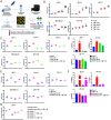
Fig. 1.
Fig. 1.
(A) Experimental workflow of the study; (B) The relative concentration of secreted particles from MCF-7 (ER+, PR+, HER2−), BT-474 (ER+, PR+, HER2+), ZR-75-1 (ER+, PR-, HER2−), MDA-MB-231 (ER−, PR−, HER2−), and MCF-10A (normal human mammary epithelial cells) obtained from NanoSight not treated and after treatment with 17β-estradiol at different concentrations (10 pM, 100 pM, 1 nM, and 10 nM); (C) The relative concentration of secreted particles from MCF-7, BT-474, ZR-75-1, MDA-MB-231, and MCF-10A obtained from NanoSight not treated and after treatment with 17β-estradiol (1 nM), 4-hydroxytamoxifen (4OHT; 100 nM), or both as normalized to the number of cells collected in each experiment; (D) Expression levels of cellular GREB1 as a marker of 17β-estradiol stimulation in MCF-7, BT-474, and ZR-75-1 cells following the treatment with 17β-estradiol (1 nM), 4-hydroxytamoxifen (4OHT, 100nM), or a combination of the two (MDA-MB-231 and MCF-10A were not tested because ER−); (E) The relative concentration of secreted particles obtained from NanoSight of MCF-7, BT-474, ZR-75-1, MDA-MB-231, and MCF-10A cell lines not treated and after treatment with 17β-estradiol (1 nM), fulvestrant (10 nM), or both, normalized to the number of cells collected in each experiment; (F) Expression levels of cellular GREB1 as a marker of 17β-estradiol stimulation in MCF-7, BT-474, and ZR-75-1 cells following the treatment with 17β-estradiol (1 nM), fulvestrant (10 nM), or both (MDA-MB-231 and MCF-10A were not tested because ER−); (G) The relative concentration of secreted particles obtained from NanoSight of MCF-7 and MDA-MB-231 cell lines after transfection with ER siRNAs and scramble control in the presence of 1 nM 17β-estradiol; FC = fold-change; ERα = estrogen receptor; 17βE = 17β-estradiol; 4-OHT = 4-hydroxytamoxifen. *P = 0.05; **P = 0.01; ***P = 0.001; ****P = 0.0001.
Fig. 2.
Fig. 2.
(A) Principal component analysis (PCA) loading displaying the clustering of the investigated sample sets used for the Affymetrix GeneChip miRNA Array 3.0; (B) Heat maps from the microarray analysis of cellular (Upper) and EV (Lower) miRNAs from MCF-7 and MDA-MB-231 with or without 17β-estradiol treatment (10 nM) highlighting the main up- or down-regulated miRNAs; (C) Venn diagrams based on the miRNA microarray data indicating the nonoverlapping differential miRNA signatures in cell and EVs between MCF-7 (red) and MDA-MB-231 (blue); (D) Validation by qRT-PCR of the expression levels of let-7a-5p (Left) and let-7d-5p (Right) in EVs and cells lines (MCF-7, BT-474, ZR-75-1, MDA-MB-231, and MCF-10A) with or without 17β-estradiol treatment (10 nM). 17βE = 17β-estradiol; FC = fold-change. *P = 0.05; **P = 0.01; ***P = 0.001; ****P = 0.0001.
Fig. 3.
Fig. 3.
(A) Expression levels of cellular miR-149-5p in MCF-7 and MDA-MB-231 following 100 pM, 1 nM, and 10 nM 17β-estradiol stimulation (three independent experiments); (B) Expression level of cellular miR-149-5p in MCF-7 and MDA-MB-231 after treatment with 10 nM 17β-estradiol, 100 nM 4-OHT, and their combination; (C) The genomic region of MIR149 from the UCSC Genomic Browser, GRCh37/hg19 with the 5 kb upstream region (“5500” promoter or p5500) containing the motif for ER1 binding; (D) Genomic area of MIR149 showing the presence of two predicted ER-binding sites (at −5,500 and −600 from the starting point of miR-149) (bottom of the figure). Chip-Seq map generated from the study of Sun et al. (49) indicating the peak in the ER-interacting region overlapping with the “5500” promoter region of miR-149-5p (extended version of the panel in SI Appendix, S3D); (E) Chip-qPCR analysis indicating the enrichment levels of the p5500 and ERE1 regions in MCF-7 and MDA-MB-231 in control and estrogen-stimulated cells following ER immunoprecipitation; (F) Diagram describing the regulatory mechanism of 17β-estradiol on miR-149-5p. FC = fold-change; 17βE = 17β-estradiol; 4-OHT = 4-hydroxytamoxifen; *P < 0.05, **P = 0.01; ***P = 0.001; ****P = 0.0001; ns = not significant.
Fig. 4.
Fig. 4.
(A) Pairing schemes of complementary miRNA–target interactions between miR-149-5p and 3′UTRs of SP1 and HNRNPA1; (B) The expression levels of SP1 and hnRNPA1 measured by western blot analysis following transfection with scrambled RNA and miR-149-5p inhibitor in MCF-7 and MDA-MB-231; (C) The expression levels of SP1 and hnRNPA1 measured by western blot analysis in MCF-7, MDA-MB-231, and MCF-10A cells not treated and after treatment with 17β-estradiol (1 nM), fulvestrant (10 nM), or both; (D) Venn diagram depicting the predicted RNA-binding proteins interacting with the let-7 family (Left) and highlighted EXO motifs for hnRNPA1 binding in the let-7a and let-7d sequences (Right) as detected with the RBP map tool; (E) Western blot analysis of EV-derived proteins from MCF-7, BT-474, ZR-75-1, MDA-MB-231, and MCF-10A cell lines, highlighting the enrichment of hnRNPA1 in the EVs (for CD63, we showed also a lower and higher exposures of membrane for a full appreciation of the presence of the protein in all cell lines); (F) Western blot analysis of hnRNPA1 biotin–streptavidin pull-down assays using biotinylated let-7a-5p and let-7d-5p probes. Input control (lane 1) indicates 20 μg total cell lysate. Lanes 2 and 3 indicate the eluted bound hnRNPA1-let-7a and -let-7d following incubation with 1 mg of total MCF-7 cellular lysate. Lane 4 corresponds to the eluted proteins resulting from the incubation of 1 mg cellular lysate with unconjugated streptavidin beads. 17βE = 17β-estradiol.
Fig. 5.
Fig. 5.
(A) Expression levels of let-7a-5p and let-7d-5p in plasma EVs (test set) from BC women stratified according to the estrogen receptor (ER) and pre/post-menopausal statuses; (B) Expression levels of let-7a-5p and let-7d-5p in serum EVs (validation set) from BC women stratified according to the ER and pre/post-menopausal statuses; (C) EV amount in the serum of ER+ BC (Upper) or TNBC patients (Lower) stratified according to BMI [BMI < 25 kg/m2 (normal weight) or BMI ≥ 25 kg/m2 (overweight and obese)]. Data are shown as median with 95% CI, Mann–Whitney U test was used to compare the medians of the two groups. miR-149-5p was also measured but was not detectable in EVs (we considered it as negative control). ns = non-significant; *P < 0.05.
Fig. 6.
Fig. 6.
(A) Workflow and experimental design of the EVs and macrophage co-culture experiment; (B) Expression levels of let-7a-5p, let-7c-5p, and let-7d-5p in THP-1 cells transfected with EVs derived from untreated MCF-7 (control), 1 nM 17β-estradiol-stimulated MCF-7 cell lines treated with scramble control RNA or with anti-let-7 probes; (C) Expression levels measured by qRT-PCR in THP-1 cells of selected cytokines as potential let-7 family regulatory targets after transfection with EVs derived from untreated MCF-7 (control), 1nM 17β-estradiol-stimulated MCF-7 cell lines treated with scramble RNA or with anti-let-7 probes; PMA = phorbol 12-myristate-13-acetate; FC = fold-change. *P = 0.05; **P = 0.01; ***P = 0.001.

References

    1. O’Brien K., Breyne K., Ughetto S., Laurent L. C., Breakefield X. O., RNA delivery by extracellular vesicles in mammalian cells and its applications. Nat. Rev. Mol. Cell Biol. 21, 585–606 (2020). - PMC - PubMed
    1. Mathieu M., Martin-Jaular L., Lavieu G., Thery C., Specificities of secretion and uptake of exosomes and other extracellular vesicles for cell-to-cell communication. Nat. Cell Biol. 21, 9–17 (2019). - PubMed
    1. Valadi H., et al. , Exosome-mediated transfer of mRNAs and microRNAs is a novel mechanism of genetic exchange between cells. Nat. Cell Biol. 9, 654–659 (2007). - PubMed
    1. Tetta C., Ghigo E., Silengo L., Deregibus M. C., Camussi G., Extracellular vesicles as an emerging mechanism of cell-to-cell communication. Endocrine 44, 11–19 (2013). - PMC - PubMed
    1. Jeppesen D. K., Zhang Q., Franklin J. L., Coffey R. J., Extracellular vesicles and nanoparticles: Emerging complexities. Trends Cell Biol. (2023), 10.1016/j.tcb.2023.01.002. - DOI - PMC - PubMed

Publication types