. 2023 Jun 6;120(23):e2221707120.
doi: 10.1073/pnas.2221707120. Epub 2023 May 30.

Steroid receptor coactivator 3 is a key modulator of regulatory T cell-mediated tumor evasion

Affiliations

Steroid receptor coactivator 3 is a key modulator of regulatory T cell-mediated tumor evasion

Sang Jun Han et al. Proc Natl Acad Sci U S A. .

Abstract

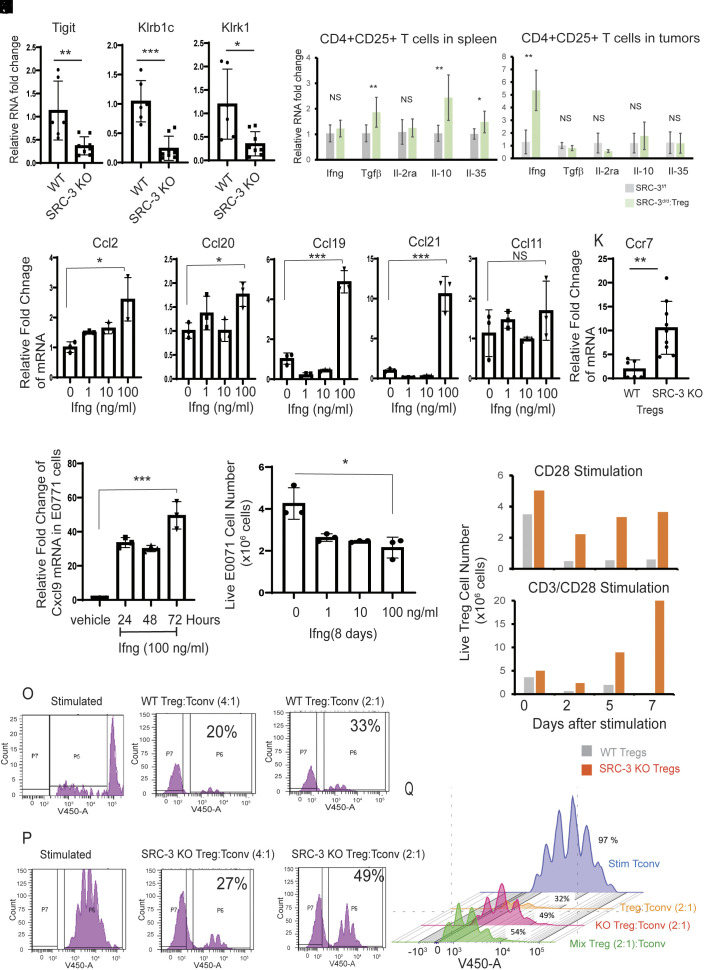
Steroid receptor coactivator 3 (SRC-3) is most strongly expressed in regulatory T cells (Tregs) and B cells, suggesting that it plays an important role in the regulation of Treg function. Using an aggressive E0771 mouse breast cell line syngeneic immune-intact murine model, we observed that breast tumors were "permanently eradicated" in a genetically engineered tamoxifen-inducible Treg-cell-specific SRC-3 knockout (KO) female mouse that does not possess a systemic autoimmune pathological phenotype. A similar eradication of tumor was noted in a syngeneic model of prostate cancer. A subsequent injection of additional E0771 cancer cells into these mice showed continued resistance to tumor development without the need for tamoxifen induction to produce additional SRC-3 KO Tregs. SRC-3 KO Tregs were highly proliferative and preferentially infiltrated into breast tumors by activating the chemokine (C-C motif) ligand (Ccl) 19/Ccl21/chemokine (C-C motif) receptor (Ccr)7 signaling axis, generating antitumor immunity by enhancing the interferon-γ/C-X-C motif chemokine ligand (Cxcl) 9 signaling axis to facilitate the entrance and function of effector T cells and natural killer cells. SRC-3 KO Tregs also show a dominant effect by blocking the immune suppressive function of WT Tregs. Importantly, a single adoptive transfer of SRC-3 KO Tregs into wild-type E0771 tumor-bearing mice can completely abolish preestablished breast tumors by generating potent antitumor immunity with a durable effect that prevents tumor reoccurrence. Therefore, treatment with SRC-3-deleted Tregs represents an approach to completely block tumor growth and recurrence without the autoimmune side effects that typically accompany immune checkpoint modulators.

Keywords: adoptive cell transfer; interferon-γ; regulatory T cells; steroid receptor coactivator 3; syngeneic murine model of breast cancer.


Conflict of interest statement

S.J.H., C.C.D., D.M.L., and B.W.O. are founding members of a new nonpublic company, called CoRegen Inc (C.C.D. and B.W.O. are Board members of CoRegen.). S.J.H., C.C.D., D.M.L., and B.W.O. have stock ownership in CoRegen. S.J.H., C.C.D., D.M.L., and B.W.O. have 4 patents issued that exist in the general area of cancer therapy. Laboratory research support exists for S.J.H., C.C.D., D.M.L., and B.W.O. from CoRegen.

Figures

Fig. 1.
Fig. 1.
E0771 breast tumor eradication in SRC-3d/d:Treg mice. (A) E0771 breast tumor growth in SRC-3f/f versus SRC-3d/d:Teg female mice. (B) Breast tumors isolated from SRC-3f/f and SRC-3d/d:Teg female mice on the 33rd day after injection with E0771 cells. (C) H&E staining of tumors is presented in panel B. (D) Spleens isolated from SRC-3f/f and SRC-3d/d:Teg female mice on the 40th day after tamoxifen treatment. (E) Breast tumor luciferase activity in SRC-3f/f and SRC-3d/d:Treg female mice. (F) The repeated tumor luciferase activity experiment with tamoxifen-treated SRC-3f/f (n = 8) and SRC-3d/d:Treg mice (n = 8) was determined on the 35th day after E0771:LUC cell injection. (G) IHC analysis of EGFP+ cells in tumors of tamoxifen-treated SRC-3f/f and SRC-3d/d:Treg mice on the 14th day after E0771 cell injection. (H) H-score for EGFP+ cells in tumors presented in panel G. (I) Tumor luciferase activity in SRC-3f/f and breast cancer–eradicated SRC-3d/d:Treg mice after a 2nd injection of E0771:LUC cells. (J) The repeated experiment for tumor luciferase activities in tamoxifen-treated SRC-3f/f (n = 5) and breast tumor–eradicated SRC-3d/d:Treg mice (n = 5) on the 35th day after E0771:LUC cell reinjection.
Fig. 2.
Fig. 2.
SRC-3d/d:Treg mice generate a tumor-suppressive immune microenvironment in breast tumors. (A) Images of tumor luciferase activity in SRC-3f/f and SRC-3d/d:Treg mice on the 3rd and 14th day after E0771:LUC cell injection. (B) H&E staining of tumors in SRC-3f/f and SRC-3d/d:Treg mice on the 14th day after E0771:LUC cell injection. (CF) IHC analysis of CD4+ T cells (C), CD8+ T cells (D), CD49b+ NK cells (E), and Foxp3+ Tregs (F) in tumors from SRC-3f/f and SRC-3d/d:Treg mice on the 14th day after E0771:LUC cell injection. The QuPath program was used to quantify IHC staining results. PLC, percentage of labeled cells.
Fig. 3.
Fig. 3.
SRC-3d/d:Treg mice possess an elevated Ifng/Cxcl9 signaling axis in breast tumors. (A) Cytokine/chemokine profiling in breast tumors obtained from SRC-3f/f and SRC-3d/d:Treg female mice on the 14th day after E0771:LUC cell injection. (B) Quantification of cytokines and chemokines shown in panel A. (C and D) IHC analysis of Ifng (C) and Cxcl9 (D) in tumors from SRC-3f/f and SRC-3d/d:Treg mice on the 14th day after E0771:LUC breast cancer cell injection using the QuPath program. (E and F) Tumor luciferase activity of tumor-bearing SRC-3d/d:Treg (E) and SRC-3f/f (F) female mice treated with anti-Ifng antibody (Ifng Ab) and control Rat IgG (Control IgG). PLC, percentage of labeled cells.
Fig. 4.
Fig. 4.
Molecular properties of SRC-3 KO Treg cells. (AC) Relative RNA levels of Tigit (A), Klrb1c (B), and Klrk1 (C) in WT versus SRC-3 KO Treg cells from spleens. (D and E) Relative mRNA levels for Ifng, Tgf-β, Il-2ra, Il-10, and Il-35 in CD4+CD25+ Treg cells isolated from spleens (D) and breast tumors (E) in breast tumor–bearing SRC-3f/f and SRC-3d/d:Treg mice on the 14th day after E0771:LUC cell injection. (FJ) Relative RNA levels of Ccl2 (F), Ccl20 (G), Ccl19(H), Ccl21(I), and Ccl11(J) in E0771 breast cancer cells treated with 0, 1, 10, and 100 ng/mL of Ifng. (K) RNA levels of Ccr7 in WT versus and SRC-3 KO Tregs. (L) RNA analysis of Cxcl9 in E0771 breast cancer cells upon Ifng (100 ng/mL) treatment for 24, 48, and 72 h. (M) The viability of E7001 breast cell numbers upon 1, 10, and 100 ng/mL of Ifng for 8 d. (N) The viability of WT Tregs and SRC-3 KO Tregs at 0, 2, 5, and 7th days after CD28 and CD3/CD28 stimulation. (O) Flow cytometry analysis of T cells showing immune suppressive activity of WT Tregs. (P) Flow cytometry analysis of T cells showing the reduced immune suppressive activity of SRC-3 KO Tregs. (Q) The reduced suppressive activity of the mixture of SRC-3 KO and WT Tregs (1:1) to Tconv cell proliferation when compared to WT Tregs. *P < 0.05; **P < 0.01; ***P < 0.001; NS, nonspecific.
Fig. 5.
Fig. 5.
Tumor eradication through adoptive SRC-3 KO Treg transfer. (A) Tumor luciferase image analysis in tumor-bearing SRC-3f/f female mice after ACT with SRC-3 KO Tregs (800K cells) and wild-type Tregs (800K cells). Control tumor-bearing SRC-3f/f mice that did not undergo ACT (NO ACT). Tumor luciferase image analysis up to the 30th day, which is in the box, and 215th day after ACT. (B) Tumor luciferase activity in SRC-3f/f female mice after ACT with different doses of SRC-3 KO Tregs. Tumor-bearing SRC-3f/f female mice were adoptively transferred with wild-type (800K cells) or SRC-3 KO Treg cells (1: 610K cells, 2: 326K cells, 3: 643K cells, and 4: 230K cells). Afterward, the luciferase activity emanating from tumors was determined. (C) Repeated ACT experiment with increased animal numbers. Tumor luciferase activity in SRC-3f/f female mice adoptively transferred with SRC-3 KO Tregs (1: 230K cells, 2: 314K cells, 3: 358K cells, and others: 800K cells, n = 9) and wild-type Tregs (800K cells, n = 9) was determined on the 28th day after E0771:LUC cell injection. (DJ) IHC analysis of EGFP+ cells (D), CD4+(E), CD8+ (F), CD49b+ (G), Foxp3 (H), Ifng (I), and Cxcl9 (J) in breast tumors of female mice at 14th day after ACT with SRC-3KO Tregs (SRC-3 KO) versus WT Tregs (Control). (K) Tumor luciferase activity in tumor-eradicated SRC-3f/f female mice after ACT with SRC-3 KO Tregs and SRC-3f/f female control mice after a 2nd injection of E0771:LUC cells.
Fig. 6.
Fig. 6.
Model for tumor eradication by SRC-3 KO Tregs. SRC-3 KO Tregs are more proliferative than WT in spleens due to increased levels of Tgf-β, Il10, and IL35 in spleens. Compared to WT Tregs, however, SRC-3 KO Tregs have reduced immune suppressive function due to the down-regulation of genes involved in immune suppression, such as TIGIT. Based on the homing activity of Tregs to cancer (Ccl19/Ccl21/Ccr7 axis) (37, 38), SRC-3 KO Tregs more aggressively infiltrate into breast tumors. In breast tumors, SRC-3 KO Tregs induced Ifng expression based on TCR activation. The elevated Ifng causes the fragility of WT Tregs in breast tumors. Also, elevated Ifng induces Cxcl9 expression in breast tumors to actively recruit cytotoxic Cxcr3+ immune cells (such as CD8+ and NK cells) into breast tumors. In addition, the recruited and tumor-resident CD8+ and NK cells activated by elevated Ifng eradicate cancer cells. The graphic was generated by BioRender.

Update of

References

    1. Dasgupta S., O’Malley B. W., Transcriptional coregulators: Emerging roles of SRC family of coactivators in disease pathology. J. Mol. Endocrinol. 53, R47–R59 (2014). - PMC - PubMed
    1. Coste A., et al. , Absence of the steroid receptor coactivator-3 induces B-cell lymphoma. The EMBO J. 25, 2453–2464 (2006). - PMC - PubMed
    1. Han S. J., Sung N., Wang J., O’Malley B. W., Lonard D. M., Steroid receptor coactivator-3 inhibition generates breast cancer antitumor immune microenvironment. Breast Cancer Res. 24, 73 (2022). - PMC - PubMed
    1. Nikolai B. C., et al. , Steroid receptor coactivator 3 (SRC-3/AIB1) is enriched and functional in mouse and human Tregs. Sci. Rep. 11, 3441 (2021). - PMC - PubMed
    1. Smith L. K., et al. , Interleukin-10 directly inhibits CD8(+) T cell function by enhancing N-glycan branching to decrease antigen sensitivity. Immunity 48, 299–312.e5 (2018). - PMC - PubMed

Publication types