. 2023 Jun 6;120(23):e2220948120.
doi: 10.1073/pnas.2220948120. Epub 2023 May 30.

Identification of broad, potent antibodies to functionally constrained regions of SARS-CoV-2 spike following a breakthrough infection

Affiliations

Identification of broad, potent antibodies to functionally constrained regions of SARS-CoV-2 spike following a breakthrough infection

Jamie Guenthoer et al. Proc Natl Acad Sci U S A. .

Abstract

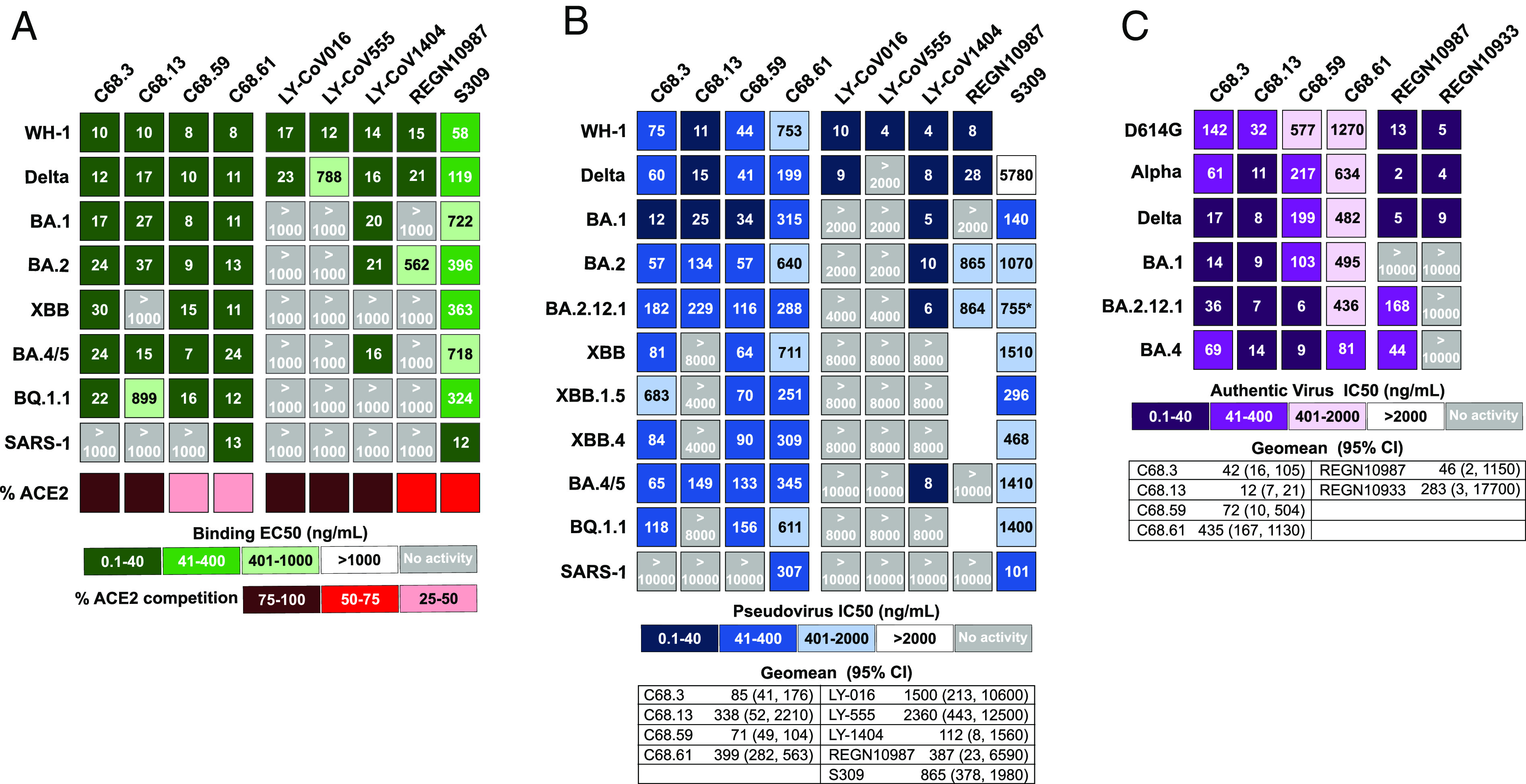
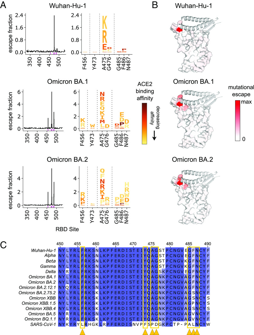
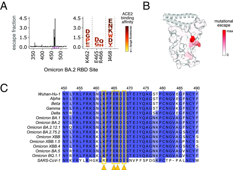
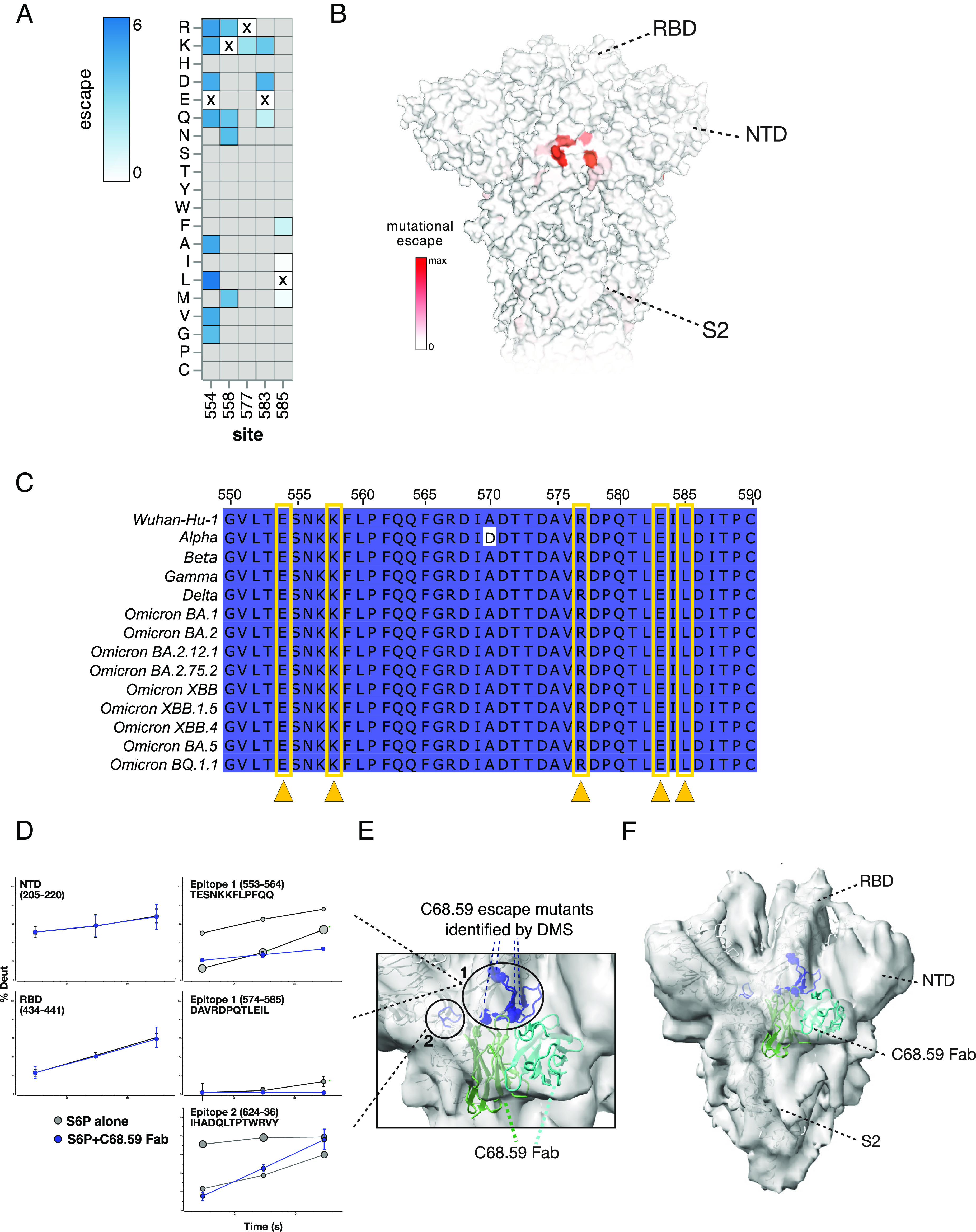
The antiviral benefit of antibodies can be compromised by viral escape especially for rapidly evolving viruses. Therefore, durable, effective antibodies must be both broad and potent to counter newly emerging, diverse strains. Discovery of such antibodies is critically important for SARS-CoV-2 as the global emergence of new variants of concern (VOC) has compromised the efficacy of therapeutic antibodies and vaccines. We describe a collection of broad and potent neutralizing monoclonal antibodies (mAbs) isolated from an individual who experienced a breakthrough infection with the Delta VOC. Four mAbs potently neutralize the Wuhan-Hu-1 vaccine strain, the Delta VOC, and also retain potency against the Omicron VOCs through BA.4/BA.5 in both pseudovirus-based and authentic virus assays. Three mAbs also retain potency to recently circulating VOCs XBB.1.5 and BQ.1.1 and one also potently neutralizes SARS-CoV-1. The potency of these mAbs was greater against Omicron VOCs than all but one of the mAbs that had been approved for therapeutic applications. The mAbs target distinct epitopes on the spike glycoprotein, three in the receptor-binding domain (RBD) and one in an invariant region downstream of the RBD in subdomain 1 (SD1). The escape pathways we defined at single amino acid resolution with deep mutational scanning show they target conserved, functionally constrained regions of the glycoprotein, suggesting escape could incur a fitness cost. Overall, these mAbs are unique in their breadth across VOCs, their epitope specificity, and include a highly potent mAb targeting a rare epitope outside of the RBD in SD1.

Keywords: SARS-CoV-2; monoclonal antibodies; spike glycoprotein; variants of concern.


Conflict of interest statement

J.O. consults for Aerium Therapeutics. T.N.S. consults for Apriori Bio on deep mutational scanning. J.D.B. serves as a scientific advisor to Apriori Bio and Oncorus. Subsequent to the completion of the research described in this manuscript, he also began to serve as a scientific advisor to Aerium Therapeutics and the Vaccine Company. H.Y.C. consults with Ellume, Merck, Abbvie, Pfizer, Medscape, Vindico, and the Bill and Melinda Gates Foundation. J.O. and J.G. are on a patent (22-173-US-PSP2) for the C68 antibodies.

Figures

Fig. 1.
Fig. 1.
Binding and neutralization measures for four C68 SARS-CoV-2 mAbs and previously authorized therapeutic mAbs. Each column in the heatmaps represents a different antibody with the C68 mAbs on the left and the therapeutic mAbs on the right. (A) Antibody binding to recombinant spike trimers from SARS-CoV-2 VOCs and SARS-CoV-1 labeled in each row. EC50 values (ng/mL), the mAb concentration that binds the spike glycoproteins at half the maximal binding signal, were calculated by nonlinear regression analysis and are the average of two independent experiments each with technical replicates. The values for the therapeutic mAbs are the average of technical replicates in one experiment. Percent inhibition of each mAb on ACE2 binding (red) assessed by competition ELISAs averaged over two independent experiments with technical replicates. (B) Neutralization of spike-pseudotyped lentiviruses by mAbs is shown. The SARS-CoV-2 VOC or SARS-CoV-1-pseudotyped viruses are labeled in each row. IC50 values (ng/mL) were calculated by nonlinear regression analysis from the average of at least three independent experiments with technical replicates tested over two different pseudotyped virus batches. Therapeutic mAbs were tested in one to two independent experiments each with two technical replicates. Geomean (95% CI) = geometric mean of the IC50s and 95% CI across SARS-CoV-2 VOCs tested (SARS-CoV-1 not included). IC50 values greater than the highest tested concentration were set to the highest concentration for this calculation. (C) Neutralization of authentic SARS-CoV-2 viruses by mAbs is shown. IC50s (ng/mL) were calculated by nonlinear regression averaged across three to four replicates in one to two independent experiments with Geomean (95% CI) = geometric mean indicated. For the EC50s and IC50s values, the smaller the value, the darker the shade, the more activity measured. Light gray boxes denote mAbs with no measured activity in the concentration range of the assay.
Fig. 2.
Fig. 2.
Profiles of mutations that escape C68.3 binding in three SARS-CoV-2 RBD libraries. (A) Line plots (Left) identify sites of binding escape (quantified as the sum of the escape fractions) at each site in the Wuhan-Hu-1 (Top), Omicron BA.1 (Middle), Omicron BA.2 (Bottom) RBDs. Escape fractions were averaged across two replicate experiments performed in independently generated libraries. Sites that have strong escape mutations (high escape fractions) in at least one of the three backgrounds are marked with pink boxes under the plots and are represented in the logo plots (Right). The logo plots show the mutations that confer escape at each of these sites where the size of the amino acid letters is scaled according to the contribution to the overall escape fraction, and the color of the mutations indicates the effect of that mutation on ACE2 binding in the specified background determined from previously published data (46, 61). A yellow mutation signifies a deleterious effect that decreases ACE2-binding affinity and dark red signifies no effect of that mutation on ACE2 binding compared to the wild-type amino acid. (B) Sites of escape for C68.3 mapped on the RBD structure (space-filled) bound to ACE2 (ribbons). The intensity of the red coloring is scaled according to the magnitude of the mutational escape fraction at each residue, with white representing no change in binding between the wild-type and mutant amino acids at that site. Sites with the highest mutation escape fractions (darkest red) indicate key binding residues in the mAb epitope. (C) Sequence alignment of a region within RBD (sites 450 to 490) across SARS-CoV-2 VOCs and SARS-CoV-1. Residues are colored based on percentage similarity at each site across the listed sequences, with darker color indicating residues invariant across the sequences. Sites with the highest escape fractions for C68.3, indicative of the binding residues across the three backgrounds, are marked with yellow arrows and boxes.
Fig. 3.
Fig. 3.
Profiles of RBD mutations that escape C68.61 in Omicron BA.2 RBD. (A) Line plot (Left) of the summed escape fraction for mutations across the Omicron BA.2 RBD sites. Escape fractions were averaged across two replicate experiments performed in independently generated libraries. The individual mutants that confer binding escape from C68.61 at those key sites are shown in the logo plots (Right) where the color of the mutations indicates the effect of that mutation on ACE2 binding (46, 61). (B) Sites of escape for C68.61 are mapped onto the RBD structure (space-filled) bound to ACE2 (ribbons). The intensity of the red coloring is scaled according to the mutational escape fraction at each residue. (C) Sequence alignment of region of RBD (WH-1 sites 450-490) across SARS-CoV-2 VOCs and SARS-CoV-1. Dark blue shows sites invariant across the sequences. Sites with the highest escape fractions for C68.61 are marked with yellow arrows and boxes. The details for this figure are as described in Fig. 2.
Fig. 4.
Fig. 4.
Mapping C68.59 epitope and neutralization escape mutations in SD1 by DMS in an Omicron BA.2 background and structural methods: HDX-MS and cryo-EM. (A) Heatmap showing the magnitude of spike-pseudotyped lentivirus neutralization escape at key sites (measured by mutation escape scores) for the mutations tested. Darker blue = more escape. Residues marked with an X are the wild-type amino acid at that site in Omicron BA.2. Gray mutations were not tested in the library. Escape scores are averaged across two replicate experiments in independently generated libraries. (B) Surface representation of spike colored by the mean escape score per site, with darker red indicating greater escape. PDB ID: 68R8 (C) Sequence alignment of region of SD1 (WH-1 sites 550 to 590) between SARS-CoV-2 VOCs, with darker color indicating residues invariant across the sequences. The key sites of escape for C68.59 are marked (yellow arrows/boxes). (DF) Structural mapping of the C68.59-binding epitope by HDX-MS and cryo-EM. (D) In response to C68.59 Fab binding, regions including the NTD and RBD do not change in their local structural ordering as reported by HDX-MS, demonstrating that those sites are not targeted by the antibody. By contrast, two adjacent sites consisting of peptide segments 553 to 564, 574 to 585, and 624 to 636 show dramatic increase in local ordering and quenching of conformational sampling when C68.59 Fab is bound to the S6P trimeric spike. Bubble plots show the uptake rates of the two distinct populations at the unbound epitope sites (gray), with the size of the bubble indicating the population fraction. (E and F) Cryo-EM reconstruction of C68.59 Fab bound to S6P structure (PDB ID 7SBP) where C68.59 Fab structure is calculated by AlphaFold fitted into map. The zoomed view of the epitope (E) and the overall architecture of the C68.59 Fab bound to S6P spike (F) are shown. Experimental electron density shown in gray, S6P structural model shown in white, C59.68 Fab shown in two shades of green to distinguish the heavy and light chains. Binding-site peptides showing protection by HDX-MS (residues 553 to 568, 575 to 585, 624 to 636) are colored blue in the cartoon structure. Residues exhibiting escape mutations by DMS are circled.

Update of

References

    1. National Institutes of Health, SARS-CoV-2 variants and susceptibility to Anti-SARS-CoV-2 monoclonal antibodies (2022). https://www.covid19treatmentguidelines.nih.gov/tables/variants-and-susce.... Accessed 6 March 2023.
    1. Collier D. A., et al. , Sensitivity of SARS-CoV-2 B.1.1.7 to mRNA vaccine-elicited antibodies. Nature 593, 136–141 (2021). - PMC - PubMed
    1. Garcia-Beltran W. F., et al. , Multiple SARS-CoV-2 variants escape neutralization by vaccine-induced humoral immunity. Cell 184, 2372–2383.e9 (2021). - PMC - PubMed
    1. Wang P., et al. , Increased resistance of SARS-CoV-2 variant P.1 to antibody neutralization. Cell Host. Microbe. 29, 747–751.e4 (2021). - PMC - PubMed
    1. Chatterjee D., et al. , SARS-CoV-2 Omicron Spike recognition by plasma from individuals receiving BNT162b2 mRNA vaccination with a 16-week interval between doses. Cell Rep. 38, 110429 (2022). - PMC - PubMed

Publication types

Supplementary concepts