

Antibody-dependent cellular cytotoxicity, infected cell binding and neutralization by antibodies to the SIV envelope glycoprotein
- PMID: 37253062
- PMCID: PMC10256149
- DOI: 10.1371/journal.ppat.1011407
Antibody-dependent cellular cytotoxicity, infected cell binding and neutralization by antibodies to the SIV envelope glycoprotein
Abstract
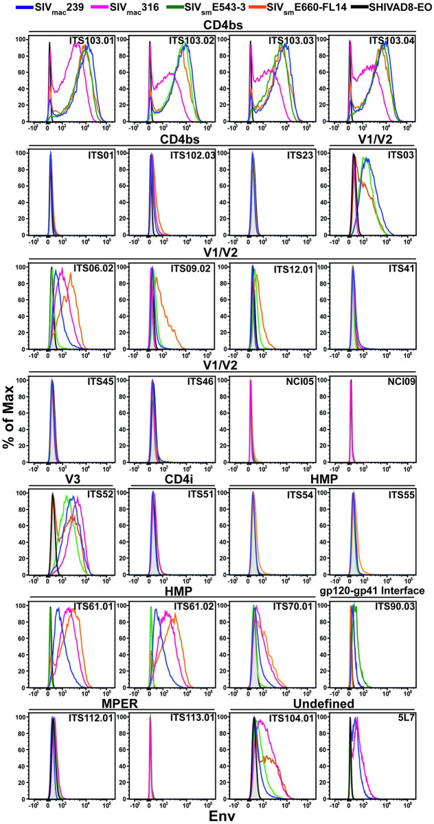
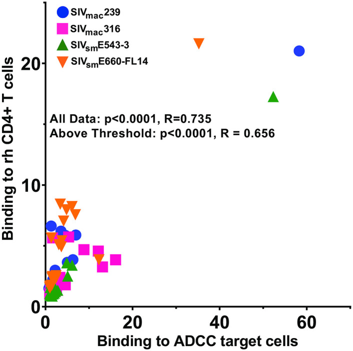
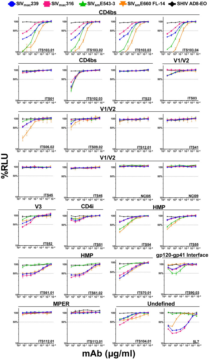
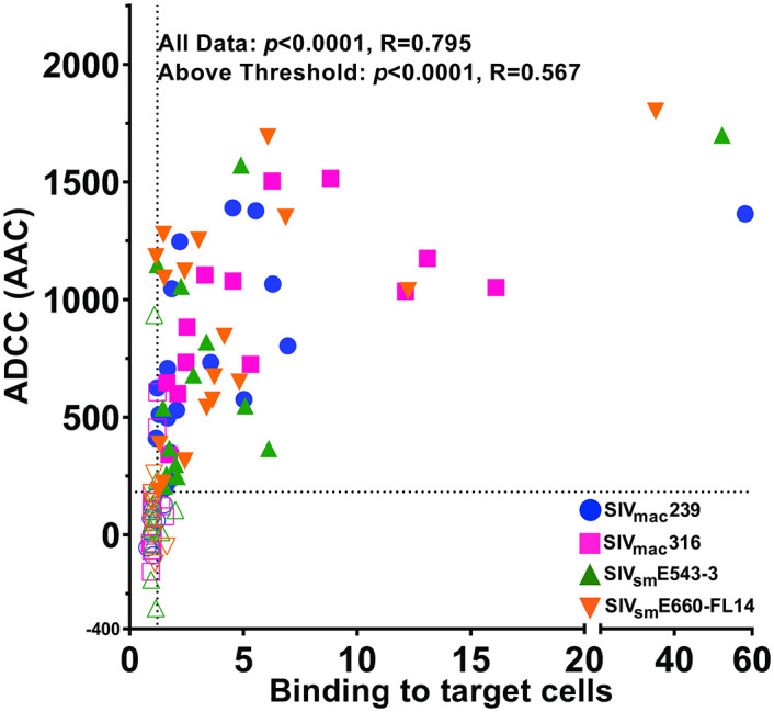
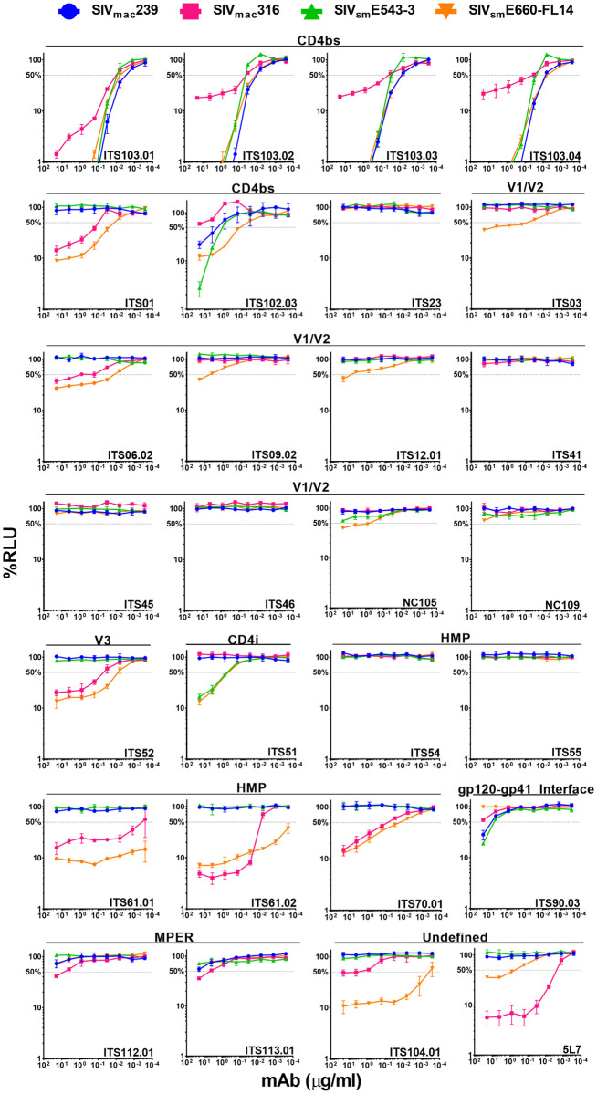
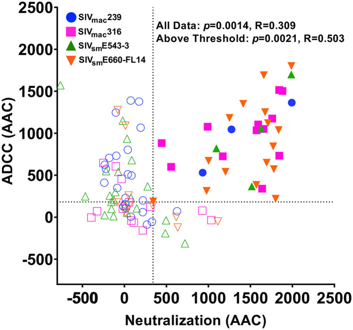
Antibodies specific for diverse epitopes of the simian immunodeficiency virus envelope glycoprotein (SIV Env) have been isolated from rhesus macaques to provide physiologically relevant reagents for investigating antibody-mediated protection in this species as a nonhuman primate model for HIV/AIDS. With increasing interest in the contribution of Fc-mediated effector functions to protective immunity, we selected thirty antibodies representing different classes of SIV Env epitopes for a comparison of antibody-dependent cellular cytotoxicity (ADCC), binding to Env on the surface of infected cells and neutralization of viral infectivity. These activities were measured against cells infected with neutralization-sensitive (SIVmac316 and SIVsmE660-FL14) and neutralization-resistant (SIVmac239 and SIVsmE543-3) viruses representing genetically distinct isolates. Antibodies to the CD4-binding site and CD4-inducible epitopes were identified with especially potent ADCC against all four viruses. ADCC correlated well with antibody binding to virus-infected cells. ADCC also correlated with neutralization. However, several instances of ADCC without detectable neutralization or neutralization without detectable ADCC were observed. The incomplete correspondence between ADCC and neutralization shows that some antibody-Env interactions can uncouple these antiviral activities. Nevertheless, the overall correlation between neutralization and ADCC implies that most antibodies that are capable of binding to Env on the surface of virions to block infectivity are also capable of binding to Env on the surface of virus-infected cells to direct their elimination by ADCC.
Copyright: This is an open access article, free of all copyright, and may be freely reproduced, distributed, transmitted, modified, built upon, or otherwise used by anyone for any lawful purpose. The work is made available under the Creative Commons CC0 public domain dedication.
Conflict of interest statement
There are no competing interests to declare.
Figures

Similar articles
-
Comparison of Antibody-Dependent Cell-Mediated Cytotoxicity and Virus Neutralization by HIV-1 Env-Specific Monoclonal Antibodies.J Virol. 2016 Jun 10;90(13):6127-6139. doi: 10.1128/JVI.00347-16. Print 2016 Jul 1. J Virol. 2016. PMID: 27122574 Free PMC article.
-
Differences in the Binding Affinity of an HIV-1 V2 Apex-Specific Antibody for the SIVsmm/mac Envelope Glycoprotein Uncouple Antibody-Dependent Cellular Cytotoxicity from Neutralization.mBio. 2019 Jul 2;10(4):e01255-19. doi: 10.1128/mBio.01255-19. mBio. 2019. PMID: 31266872 Free PMC article.
-
A novel assay for antibody-dependent cell-mediated cytotoxicity against HIV-1- or SIV-infected cells reveals incomplete overlap with antibodies measured by neutralization and binding assays.J Virol. 2012 Nov;86(22):12039-52. doi: 10.1128/JVI.01650-12. Epub 2012 Aug 29. J Virol. 2012. PMID: 22933282 Free PMC article.
-
Beyond Viral Neutralization.AIDS Res Hum Retroviruses. 2017 Aug;33(8):760-764. doi: 10.1089/AID.2016.0299. Epub 2017 Feb 16. AIDS Res Hum Retroviruses. 2017. PMID: 28084796 Free PMC article. Review.
-
Epitope specificity of human immunodeficiency virus-1 antibody dependent cellular cytotoxicity [ADCC] responses.Curr HIV Res. 2013 Jul;11(5):378-87. doi: 10.2174/1570162x113116660059. Curr HIV Res. 2013. PMID: 24191939 Free PMC article. Review.
Cited by
-
AZD5582 plus SIV-specific antibodies reduce lymph node viral reservoirs in antiretroviral therapy-suppressed macaques.Nat Med. 2023 Oct;29(10):2535-2546. doi: 10.1038/s41591-023-02570-7. Epub 2023 Oct 2. Nat Med. 2023. PMID: 37783968 Free PMC article.
-
Combining a rhesus cytomegalovirus/SIV vaccine with a neutralizing antibody to protect against SIV challenges in rhesus macaques.Front Microbiol. 2025 Jun 2;16:1592647. doi: 10.3389/fmicb.2025.1592647. eCollection 2025. Front Microbiol. 2025. PMID: 40529575 Free PMC article.
-
Adeno-associated viral delivery of Env-specific antibodies prevents SIV rebound after discontinuing antiretroviral therapy.bioRxiv [Preprint]. 2024 Jun 3:2024.05.30.593694. doi: 10.1101/2024.05.30.593694. bioRxiv. 2024. Update in: Sci Immunol. 2025 Feb 28;10(104):eadq4973. doi: 10.1126/sciimmunol.adq4973. PMID: 38895320 Free PMC article. Updated. Preprint.
-
Potent antibody-dependent cellular cytotoxicity of a V2-specific antibody is not sufficient for protection of macaques against SIV challenge.PLoS Pathog. 2024 Jan 22;20(1):e1011819. doi: 10.1371/journal.ppat.1011819. eCollection 2024 Jan. PLoS Pathog. 2024. PMID: 38252675 Free PMC article.
-
Potent broadly neutralizing antibodies mediate efficient antibody-dependent phagocytosis of HIV-infected cells.PLoS Pathog. 2024 Oct 28;20(10):e1012665. doi: 10.1371/journal.ppat.1012665. eCollection 2024 Oct. PLoS Pathog. 2024. PMID: 39466835 Free PMC article.
References
Publication types
MeSH terms
Substances
Grants and funding
LinkOut - more resources
Full Text Sources
Medical
Research Materials
